
Клинический разбор в общей медицине №03 2026
The mechanobiology of Alzheimer’s disease: PIEZO1 as the nexus of brain stiffening and neurodegeneration
Номера страниц в выпуске:11-14
Abstract
Background. Alzheimer’s disease (AD) pathogenesis extends beyond biochemical alterations to include biomechanical dysfunction. Brain tissue stiffening, vascular shear stress changes, and impaired fluid dynamics characterize the AD brain. PIEZO1, a mechanosensitive ion channel, translates these mechanical cues into pathological signaling across neural cells.
Objective: This review synthesizes current evidence on PIEZO1 in AD, proposing an integrated mechanobiological model in which PIEZO1 links brain mechanical remodeling to cellular and molecular pathologies.
Methods. A comprehensive literature search of PubMed and Scopus was conducted to identify studies on PIEZO1 expression, activation, and downstream signaling in the context of AD. Molecular, cellular, and animal studies involving microglia, astrocytes, neurons, endothelial cells, and pericytes were critically evaluated to construct a unified model of mechanotransductive dysfunction.
Results. PIEZO1 dysfunction affects multiple systems in AD. Vascular impairment reduces shear-stress responses, causing hypoperfusion and blood-brain barrier leakage. Glymphatic clearance is compromised via reduced vascular pulsatility, weakening Aβ removal. Microglial PIEZO1 senses Aβ stiffness to promote early phagocytosis, yet chronic activation drives neuroinflammation; astrocytic PIEZO1 contributes to reactive gliosis. In neurons, abnormal PIEZO1 signaling disrupts synaptic plasticity. This establishes a self-reinforcing cycle: tissue stiffening further activates PIEZO1, exacerbating pathology.
Conclusion. PIEZO1 is a central mechanotransducer, integrating biomechanical alterations into AD pathogenesis. PIEZO1 emerges as a high-value, yet complex, therapeutic target in AD. Realizing its potential will depend on the development of precision 'mechanopharmacology'—interventions that are not only cell- and stage-specific but also capable of fine-tuning channel activity rather than simply activating or inhibiting it. Future studies must prioritize the development of targeted modulators and a deeper understanding of the temporal dynamics of PIEZO1 signaling across the disease continuum.
Keywords: PIEZO1, mechanobiology, Alzheimer’s disease, neurovascular unit, tissue stiffness, calcium signaling.
For citation: Khaled A. Abdel-Sater. The mechanobiology of Alzheimer’s disease: PIEZO1 as the nexus of brain stiffening and neurodegeneration. Clinical review for general practice. 2026; 7 (3): 11–14. DOI: 10.47407/kr2026.7.3.00785
Механобиология болезни Альцгеймера: PIEZO1 как связующее звено между снижением упругости мозговой ткани и процессом отмирания нейронов
Халед А. Абдель-СатерУниверситет Мута, Эль-Карак, Иордания
Kabdelsater@mutah.edu.jo
Аннотация
Актуальность. Патогенез болезни Альцгеймера (БА) выходит за рамки биохимических изменений. Он включает в себя биомеханическую дисфункцию. При БА головной мозг характеризуется снижением упругости мозговой ткани, изменением напряжения сдвига стенок сосудов и нарушением динамики циркуляции жидкости. Механочувствительный ионный канал PIEZO1 преобразует эти механические сигналы в патологические сигналы, передаваемые нервными клетками.
Цель. В обзоре обобщены имеющиеся данные по PIEZO1 при БА, предложена комплексная механобиологическая модель, в которой PIEZO1 связывает механическое ремоделирование головного мозга с нарушениями на клеточном и молекулярном уровнях.
Методы. Выполнен поиск литературных источников в PubMed и Scopus с целью идентифицировать исследования экспрессии, активации и дальнейшей передачи сигнала PIEZO1 в контексте БА. Проведена критическая оценка исследований микроглии, астроцитов, нейронов, эндотелиальных клеток и перицитов на молекулярном и клеточном уровне, а также на животных с целью создать единую модель механотрансдуктивной дисфункции.
Результаты. При БА дисфункция PIEZO1 влияет на многочисленные системы организма. Сосудистые нарушения приводят к уменьшению реакции на изменение напряжения сдвига, вызывая гипоперфузию и повреждение гематоэнцефалического барьера. Вследствие уменьшения пульсации сосудов нарушается глимфатический клиренс, из-за чего хуже выводится Aβ. PIEZO1 в микроглии определяет связанное с Aβ снижение упругости, способствуя началу фагоцитоза, однако его постоянное пребывание в активном состоянии вызывает нейровоспаление; PIEZO1 в астроцитах способствует развитию реактивного глиоза. В нейронах нарушение PIEZO1 сигнализации приводит к утрате синаптической пластичности. Таким образом, устанавливается самоусиливающийся цикл: снижение упругости тканей обеспечивает все большую активность PIEZO1, приводя к обострению заболевания.
Выводы. PIEZO1 представляет собой основной механотрансдуктор, обеспечивающий интеграцию биомеханических изменений в патогенез БА. При БА PIEZO1 выступает в роли чрезвычайно значимой, но сложной терапевтической мишени. Реализация его потенциала будет зависеть от развития прецизионной «механофармакологии» – вмешательств, которые не только специфичны для определенных клеток и стадий, но также способны обеспечивать тонкую настройку активности каналов, а не просто активировать или ингибировать их. В ходе дальнейших исследований приоритет должен быть отдан разработке целевых модуляторов и более глубокому изучению временной динамики PIEZO1 сигнализации на протяжении всего периода болезни.
Ключевые слова: PIEZO1, механобиология, болезнь Альцгеймера, нейрососудистая единица, снижение упругости ткани, кальциевая сигнализация.
Для цитирования: Халед А. Абдель-Сатер. Механобиология болезни Альцгеймера: PIEZO1 как связующее звено между снижением упругости мозговой ткани и процессом отмирания нейронов. Клинический разбор в общей медицине. 2026; 7 (3): 11–14. DOI: 10.47407/kr2026.7.3.00785
Introduction
Alzheimer’s disease (AD) is classically defined by amyloid-β (Aβ) plaques, tau tangles, and neuroinflammation [1]. Beyond biochemistry, the brain’s mechanical properties – extracellular matrix (ECM) stiffness, vascular shear stress, and interstitial pressure – are dynamically regulated. Aging and AD alter these properties: protein aggregation and ECM remodeling stiffen parenchyma [1, 2], while cerebral vessels lose compliance, impairing perfusion [3].
Mechanobiology – the study of how cells sense and respond to mechanical forces—offers a new perspective on AD pathogenesis. CNS cells express mechanosensitive ion channels that translate physical cues into biochemical signals. PIEZO1, a non-selective cation channel, is widely expressed in microglia, astrocytes, neurons, and endothelial cells and is highly sensitive to membrane tension, substrate stiffness, and shear stress [4].
A growing body of evidence directly implicates PIEZO1 in AD pathology. Microglial PIEZO1 senses the heightened stiffness of Aβ fibrils, triggering Ca2+ influx that promotes phagocytosis and plaque clearance; its loss exacerbates plaque burden and cognitive deficits in mice [5]. Pharmacological activation of PIEZO1 with agonists like Yoda1 enhances microglial survival, phagocytosis, and Aβ clearance in human models, directly linking channel activity to plaque reduction [6]. Similarly, astrocytes surrounding amyloid plaques upregulate PIEZO1, particularly under inflammatory conditions [7]. This positions PIEZO1 as an active sensor of the altered mechanical landscape of the AD brain, potentially driving key pathological processes.
This review argues that PIEZO1 is not merely a passive sensor but a central integrator and amplifier. It converts the brain's physical deterioration into biochemical and cellular dysfunction, which in turn promotes further mechanical remodeling, creating a self-reinforcing pathogenic cycle that accelerates disease progression. We propose a unified mechanobiological framework for AD, in which PIEZO1 dysfunction connects vascular deficits, glial activation, and synaptic failure into a cohesive model of neurodegeneration.
Structure and mechanogating of PIEZO1
PIEZO1 is a large, trimeric ion channel with a unique three-bladed "propeller" architecture. Each subunit forms a curved blade that radiates from a central pore, creating a distinctive nano-dome structure within the membrane. Cryo-electron microscopy studies have identified intracellular "beam-like" elements that tether the blades to the pore, functioning as levers to transmit mechanical force into channel gating [8]. Upon activation by stimuli such as membrane stretch or changes in substrate stiffness, PIEZO1 opens, permitting a rapid influx of cations, primarily Ca2+ and Na+ [9]. This ion flux initiates diverse downstream signaling cascades, including the activation of calcium/calmodulin-dependent kinases (CaMKs), calcineurin, NF-κB, and triggers cytoskeletal reorganization and transcriptional changes [10]. Research tools like the agonist Yoda1 and the peptide antagonist GsMTx4 are instrumental in probing PIEZO1's functions [9]. Although its gating kinetics are cell-type-dependent, the fundamental principle of force-to-signal conversion is a conserved feature of PIEZO1 biology [10].
Mechanical remodeling in the Alzheimer’s brain
The AD brain undergoes significant mechanical remodeling that actively drives disease progression [11]. Tissue stiffness escalates due to the combined effects of Aβ plaques, neurofibrillary tangles, and reactive gliosis, which promote the deposition of ECM proteins like collagen [11, 12]. Concurrently, vascular mechanics are compromised, featuring reduced cerebral blood flow, arterial stiffening, and blood-brain barrier (BBB) breakdown, which disrupts shear stress and interstitial fluid dynamics [13].
PIEZO1 is aberrantly activated in this altered mechanical environment. For instance, microglia encountering stiff Aβ plaques upregulate PIEZO1, initiating Ca2+-dependent signaling pathways [11]. This mechanosensitive activation extends across cell types: glial PIEZO1 promotes neuroinflammation via NF-κB and inflammasomes [14]; endothelial PIEZO1 modulates BBB integrity and oxidative stress [15]; and neuronal PIEZO1 influences Ca2+ homeostasis and potentially tau pathology [14].
This creates a self-reinforcing, feed-forward cycle. Initial Aβ deposition and aging-related stiffening activate PIEZO1, which in turn amplifies vascular dysfunction, impairs glymphatic clearance, heightens inflammation, and promotes further ECM remodeling and neuronal injury [16, 17]. This mechanopathogenic cycle establishes PIEZO1 as a central node, physically linking the brain's deteriorating biomechanics to the classic hallmarks of AD [15, 17] (Figure).
Cell-type-specific roles of PIEZO1 in ad pathobiology
Microglia: Sensing Stiffness and Directing Amyloid Clearance. Microglial PIEZO1, upregulated near Aβ plaques, senses fibril stiffness, triggering Ca2+ influx that drives clustering, migration, and phagocytic clearance of Aβ. The functional importance of this pathway is underscored by evidence that microglial PIEZO1 deletion in mice increases plaque burden and accelerates cognitive decline, whereas pharmacological activation with Yoda1 enhances lysosomal activity, Aβ phagocytosis, and memory [5, 6].
Astrocytes: Modulating Reactive States and Synaptic Function. Reactive astrocytes surrounding amyloid plaques exhibit heightened PIEZO1 expression, a response amplified by peripheral inflammation [7]. Activation of astrocytic PIEZO1 regulates Ca2+ oscillations, ATP release, and cytokine profiles, often reducing pro-inflammatory factors like IL-1β and TNF-α [18]. In vivo, astrocyte-specific PIEZO1 deletion disrupts hippocampal neurogenesis, long-term potentiation (LTP), and memory, while its overexpression enhances synaptic plasticity and cognition [19]. The detrimental role of PIEZO1 in inflammation is further amplified by the finding that inflammatory conditions can 'sensitize' astrocytic PIEZO1 channels, lowering their activation threshold and leading to exaggerated calcium responses that fuel neuroinflammation.
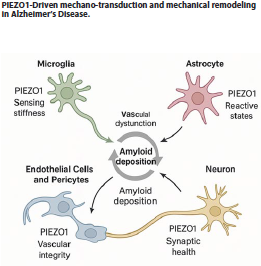 Endothelial Cells and Pericytes: Governing Vascular Integrity
As a primary shear-stress sensor in the vasculature, endothelial PIEZO1 generates Ca2+ signals in brain capillaries, implicating it in neurovascular coupling [20]. Pathologically, Aβ1-40 directly potentiates PIEZO1 activity in endothelial cells, increasing its mechanosensitivity [17]. Furthermore, endothelial PIEZO1 mediates blood-flow-dependent pericyte proliferation via Notch signaling, a key process for neurovascular unit development and integrity [22]. In disease states, however, excessive PIEZO1 activation can contribute to BBB disruption and inflammation, highlighting its dual role in vascular health and pathology. The critical role of PIEZO1 in vascular pathology is underscored by in vivo evidence showing that its inhibition with the antagonist GsMTx4 preserves BBB integrity and prevents cognitive impairment in models of chronic cerebral hypoperfusion, a key contributor to vascular dementia.
Neurons: Direct and Indirect Influences on Synaptic Health
While PIEZO1 expression is lower in mature neurons than in glia, it can influence neurite outgrowth, synaptic plasticity, and Ca2+ homeostasis [4]. More significantly, PIEZO1 indirectly governs neuronal health through its roles in microglial plaque clearance, astrocytic synaptic support, and endothelial regulation of blood flow. Preliminary evidence also suggests a potential link to tau phosphorylation, meriting further investigation.
The context-dependent duality of PIEZO1 signaling
The impact of PIEZO1 activation is not universally beneficial or detrimental but is instead determined by cell type, disease stage, and pathological context.
• Microglia: In early AD, PIEZO1 activation is protective, promoting Aβ phagocytosis [5]. In chronic phases, sustained activation may lead to oxidative stress and phagocytic dysfunction [11].
• Vasculature: Under physiological conditions, PIEZO1 is crucial for flow sensing and vascular development [22]. In chronic hypoperfusion or advanced AD, its overactivation contributes to BBB disruption by driving oxidative stress and tight junction damage via the CaMKII/Nrf2 pathway [15]. Therapeutically, PIEZO1 inhibition with GsMTx4 in such contexts has been shown to preserve BBB integrity, reduce neuroinflammation, and prevent cognitive impairment [23].
• Astrocytes: Acute PIEZO1 signaling supports neurogenesis and synaptic function [19]. Under persistent mechanical stress or inflammation, it may drive the transition to a chronic reactive state, exacerbating gliosis [24].
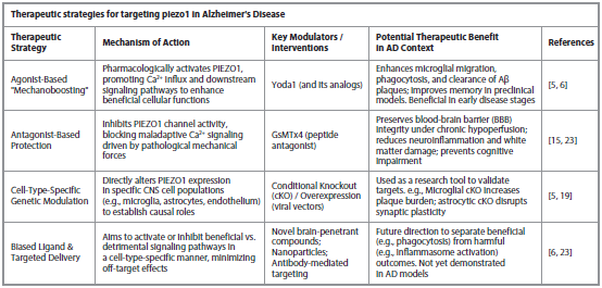
Mechanopharmacology: therapeutic modulation of PIEZO1
The dual nature of PIEZO1 signaling necessitates precise therapeutic strategies.
• Agonist-Based "Mechanoboosting": Using compounds like Yoda1 to activate microglial PIEZO1 represents a promising strategy to enhance Aβ clearance in early AD, as demonstrated in preclinical models [6].
• Antagonist-Based Protection: In contexts of vascular dysfunction, inhibitors like GsMTx4 can block maladaptive PIEZO1 signaling, protecting the BBB and reducing inflammation [23].
• Challenges and Future Directions: The primary challenge is achieving cell-type and context specificity to avoid off-target effects, given PIEZO1's widespread expression. Future efforts must focus on developing brain-penetrant, biased modulators, and targeted delivery systems (e.g., nanoparticles, viral vectors) to direct therapies to specific cell populations.
Furthermore, the potential benefits of PIEZO1 inhibition may extend beyond the vasculature. Evidence suggests that maladaptive PIEZO1 signaling contributes to white matter damage, as GsMTx4 has been shown to attenuate demyelination in certain models. This opens another promising avenue for therapeutic intervention in AD, where white matter integrity is frequently compromised (Table).
Research frontiers and key gaps
To translate these mechanistic insights into therapies, several priorities must be addressed:
1. Conditional Genetic Models. Cell-type-specific (e.g., microglia, astrocyte, endothelium) PIEZO1 knockout/overexpression studies in AD models are needed to establish causal relationships.
2. Temporal Dynamics. Longitudinal studies mapping PIEZO1 expression and activity across disease stages are crucial to determine the optimal window for intervention.
3. Downstream Signaling. A comprehensive mapping of PIEZO1-triggered pathways (CaMK, NF-κB, NLRP3) in each CNS cell type in AD contexts is required.
4. Human Validation. Investigating PIEZO1 expression and activation in human post-mortem AD brain tissue using spatial transcriptomics and proteomics is essential.
5. Drug Development. There is an urgent need for novel, brain-penetrant PIEZO1 modulators. Future efforts should focus on developing biased ligands that can activate beneficial downstream pathways (e.g., for phagocytosis) while avoiding detrimental ones (e.g., for inflammasome activation), as well as advanced delivery systems (e.g., nanoparticle – or antibody-mediated targeting) for cell-type-specific modulation.
Conclusion
PIEZO1 functions as a central mechanotransducer in AD, linking pathological brain stiffening to signaling pathways that regulate Aβ clearance, neuroinflammation, vascular integrity, and synaptic function. Its dual, context-dependent roles are both a challenge and opportunity. Realizing its therapeutic potential requires precision mechanopharmacology – interventions that are molecule-, cell-, and stage-specific. Incorporating biomechanical perspectives, targeting PIEZO1 provides a novel, holistic approach to disrupt neurodegeneration and alter disease progression.
Conflict of interests. The author declares that there is not conflict of interests.
Конфликт интересов. Автор заявляет об отсутствии конфликта интересов.
Список литературы доступен на сайте журнала https://klin-razbor.ru/
The list of references is available on the journal‘s website https://klin-razbor.ru/
Информация об авторе
Information about the author
Khaled A. Abdel-Sater – MD, Department of Dental and Medical Sciences, Faculty of Dentistry, Mutah University, Alkarak, Jordan. E-mail: Kabdelsater@mutah.edu.jo; ORCID: 0000-0001-9357-4983
Халед А. Абдель-Сатер – доктор медицины, Кафедра стоматологии и медицинских наук, стоматологический факультет, Университет Мута, Эль-Карак, Иордания.
E-mail: Kabdelsater@mutah.edu.jo; ORCID: 0000-0001-9357-4983
Поступила в редакцию: 13.11.2025
Поступила после рецензирования: 24.11.2025
Принята к публикации: 04.12.2025
Received: 13.11.2025
Revised: 24.11.2025
Accepted: 04.12.2025
Список исп. литературыСкрыть список1. Lepelletier FX, Mann DM, Robinson AC et al. Early changes in extracellular matrix in Alzheimer's disease. Neuropathol Appl Neurobiol 2017;43(2):167-82.
2. Murphy MC, Jones DT, Jack CR Jr et al. Regional brain stiffness changes across the Alzheimer's disease spectrum. Neuroimage Clin 2015;10:283-90.
3. Liu X, Halvorsen S, Blanke N et al. Progressive mechanical and structural changes in anterior cerebral arteries with Alzheimer's disease. Alzheimers Res Ther 2023;15(1):185.
4. Zheng Q, Liu H, Yu W et al. Mechanical properties of the brain: Focus on the essential role of Piezo1-mediated mechanotransduction in the CNS. Brain Behav 2023;13(9):e3136.
5. Hu J, Chen Q, Zhu H et al. Microglial Piezo1 senses Aβ fibril stiffness to restrict Alzheimer's disease. Neuron 2023;111(1):15-29.e8.
6. Jäntti H, Sitnikova V, Ishchenko Y et al. Microglial amyloid beta clearance is driven by PIEZO1 channels. J Neuroinflammation 2022;19(1):147.
7. Velasco-Estevez M, Mampay M, Boutin H et al. Infection Augments Expression of Mechanosensing Piezo1 Channels in Amyloid Plaque-Reactive Astrocytes. Front Aging Neurosci 2018;10:332.
8. Guo YR, MacKinnon R. Structure-based membrane dome mechanism for Piezo mechanosensitivity. Elife 2017;6:e33660.
9. Zhao Q, Zhou H, Li X, Xiao B. The mechanosensitive Piezo1 channel: a three-bladed propeller-like structure and a lever-like mechanogating mechanism. FEBS J 2019;286(13):2461-70.
10. Liu Y, Xu YQ, Long YY et al. Mechanosensitive channel Piezo1 in calcium dynamics: structure, function, and emerging therapeutic strategies. Front Mol Biosci 2025;12:1693456.
11. Liu Y, Zhang J, Zhao Y et al. Mechanotransduction Activates Microglia and Impairs Phagocytosis in Stiff Amyloid-β Plaques. Adv Sci (Weinh) 2025;12(30):e03389.
12. Bangen KJ, Smirnov DS, Delano-Wood L et al. Arterial stiffening acts synergistically with APOE genotype and AD biomarker status to influence memory in older adults without dementia. Alzheimers Res Ther 2021;13(1):121.
13. Yang B, Li Z, Li P et al. Piezo1 in microglial cells: Implications for neuroinflammation and tumorigenesis. Channels (Austin) 2025;19(1):2492161.
14. Fu H, Yu Y, Wang S et al. Piezo1 disrupts blood-brain barrier via CaMKII/Nrf2 in ischemic stroke. Cell Mol Life Sci 2025;82(1):259.
15. Pirri C. PIEZO Channels in Mechano-Inflammation: Gatekeepers of Neuroimmune Crosstalk. Diseases 2025;13(8):263.
16. Lim XR, Willemse L, Harraz OF. Amyloid beta Aβ1-40 activates Piezo1 channels in brain capillary endothelial cells. Biophys J 2024;124(24): 4432-41.
17. Velasco-Estevez M, Rolle SO, Mampay M et al. Piezo1 regulates calcium oscillations and cytokine release from astrocytes. Glia 2020;68(1):145-60.
18. Chi S, Cui Y, Wang H et al. Astrocytic Piezo1-mediated mechanotransduction determines adult neurogenesis and cognitive functions. Neuron 2022;110(18):2984-99.e8.
19. Harraz OF, Klug NR, Senatore AJ et al. Piezo1 Is a Mechanosensor Channel in Central Nervous System Capillaries. Circ Res 2022;130(10):1531-46.
20. Zi H, Peng X, Cao J et al. Piezo1-dependent regulation of pericyte proliferation by blood flow during brain vascular development. Cell Rep 2024;43(1):113652.
21. Xu F, Xin Q, Ren M et al. Inhibition of piezo1 prevents chronic cerebral hypoperfusion-induced cognitive impairment and blood brain barrier disruption. Neurochem Int 2024;175:105702.
22. Yu D, Ahmed A, Jayasi J et al. Inflammation condition sensitizes Piezo1 mechanosensitive channel in mouse cerebellum astrocyte. Front Cell Neurosci 2023;17:1200946.
23. Xu F, Xin Q, Ren M, Shi P, Wang B. Inhibition of piezo1 prevents chronic cerebral hypoperfusion-induced cognitive impairment and blood brain barrier disruption. Neurochem Int. 2024;175:105702. DOI:10.1016/j.neuint.2024.105702