


Клинический разбор в общей медицине №07 2026
Совпадение маркеров аутоиммунных заболеваний при шистосомозе печени: клинический случай
Номера страниц в выпуске:103-106
Аннотация
Введение. Шистосомоз – это паразитарное заболевание, поражающее гепатобилиарную систему, которое может напоминать аутоиммунное заболевание или хроническую печеночную недостаточность. Его клинические проявления могут быть такими же, как при аутоиммунном гепатите или первичном билиарном холангите, особенно у лиц из эндемичных регионов. Для постановки диагноза обычно требуются серологические исследования, исследования методами медицинской визуализации и гистопатологические исследования. Представлен случай шистосомоза печени у мужчины среднего возраста, обратившегося с жалобами на хроническую боль в правом верхнем квадранте живота, для которого были получены неоднозначные результаты серологических исследований маркеров аутоиммунных заболеваний.
Описание клинического случая. Мужчина в возрасте 42 лет, не имевший серьезных заболеваний или хирургических вмешательств в анамнезе, обратился 25 апреля 2025 г. с жалобами на периодические ночные боли в правом верхнем квадранте живота и сниженный аппетит. Первоначальные результаты лабораторных исследований показали сохранную функцию печени (АЛТ 29 Ед/л, АСТ 28 Ед/л, щелочная фосфатаза 84 Ед/л, общий билирубин 11,4 мкмоль/л), нормальные уровни маркеров аутоиммунных заболеваний (СРБ
1,7 мг/л) и наличие IgG к Schistosoma (ИФА). Углубленное исследование маркеров аутоиммунных заболеваний выявило положительные результаты для AMA-M2 (33,4 Ед) и ASMA (31 Ед), а также пограничные результаты для АНА (1:100). Результаты ультразвукового исследования брюшной полости и FibroScan (11,3 кПа) позволили предположить наличие фиброза; МРТ, выполненная 1 мая 2025 г., выявила неровные края печени без очаговых поражений. Результаты исследования кала на яйца гельминтов были отрицательными. Биопсия печени, выполненная 23 июня 2025 г., подтвердила шистосомоз печени и небольшое воспаление воротной вены. Пациента направили в инфекционную больницу для проведения противопаразитарной терапии 3 июля 2025 г.
Заключение. Представленный случай демонстрирует шистосомоз печени, проявляющийся совпадением маркеров аутоиммунных заболеваний, которое затрудняет диагностику. У пациентов из эндемичных районов паразитарные инфекции следует рассматривать в качестве дифференциального диагноза даже при наличии аутоантител. Необходимо длительное наблюдение для отслеживания совпадения маркеров аутоиммунных заболеваний и прогрессирования фиброза печени.
Ключевые слова: шистосомоз печени, маркеры аутоиммунных заболеваний, боль в правом верхнем квадранте живота, фиброз печени, биопсия печени.
Для цитирования: Равия Мубарак Мохамед, Ашраф Альаккад, Махер Вассим Малаиб. Совпадение маркеров аутоиммунных заболеваний при шистосомозе печени: клинический случай. Клинический разбор в общей медицине. 2025; 7 (6): 103–106. DOI: 10.47407/kr2026.7.7.00893
Введение. Шистосомоз – это паразитарное заболевание, поражающее гепатобилиарную систему, которое может напоминать аутоиммунное заболевание или хроническую печеночную недостаточность. Его клинические проявления могут быть такими же, как при аутоиммунном гепатите или первичном билиарном холангите, особенно у лиц из эндемичных регионов. Для постановки диагноза обычно требуются серологические исследования, исследования методами медицинской визуализации и гистопатологические исследования. Представлен случай шистосомоза печени у мужчины среднего возраста, обратившегося с жалобами на хроническую боль в правом верхнем квадранте живота, для которого были получены неоднозначные результаты серологических исследований маркеров аутоиммунных заболеваний.
Описание клинического случая. Мужчина в возрасте 42 лет, не имевший серьезных заболеваний или хирургических вмешательств в анамнезе, обратился 25 апреля 2025 г. с жалобами на периодические ночные боли в правом верхнем квадранте живота и сниженный аппетит. Первоначальные результаты лабораторных исследований показали сохранную функцию печени (АЛТ 29 Ед/л, АСТ 28 Ед/л, щелочная фосфатаза 84 Ед/л, общий билирубин 11,4 мкмоль/л), нормальные уровни маркеров аутоиммунных заболеваний (СРБ
1,7 мг/л) и наличие IgG к Schistosoma (ИФА). Углубленное исследование маркеров аутоиммунных заболеваний выявило положительные результаты для AMA-M2 (33,4 Ед) и ASMA (31 Ед), а также пограничные результаты для АНА (1:100). Результаты ультразвукового исследования брюшной полости и FibroScan (11,3 кПа) позволили предположить наличие фиброза; МРТ, выполненная 1 мая 2025 г., выявила неровные края печени без очаговых поражений. Результаты исследования кала на яйца гельминтов были отрицательными. Биопсия печени, выполненная 23 июня 2025 г., подтвердила шистосомоз печени и небольшое воспаление воротной вены. Пациента направили в инфекционную больницу для проведения противопаразитарной терапии 3 июля 2025 г.
Заключение. Представленный случай демонстрирует шистосомоз печени, проявляющийся совпадением маркеров аутоиммунных заболеваний, которое затрудняет диагностику. У пациентов из эндемичных районов паразитарные инфекции следует рассматривать в качестве дифференциального диагноза даже при наличии аутоантител. Необходимо длительное наблюдение для отслеживания совпадения маркеров аутоиммунных заболеваний и прогрессирования фиброза печени.
Ключевые слова: шистосомоз печени, маркеры аутоиммунных заболеваний, боль в правом верхнем квадранте живота, фиброз печени, биопсия печени.
Для цитирования: Равия Мубарак Мохамед, Ашраф Альаккад, Махер Вассим Малаиб. Совпадение маркеров аутоиммунных заболеваний при шистосомозе печени: клинический случай. Клинический разбор в общей медицине. 2025; 7 (6): 103–106. DOI: 10.47407/kr2026.7.7.00893
Clinical Case
Rawia Mubarak Mohamed1, Ashraf ALakkad2, Maher Wassim Malaeb1
1 Burjeel Medical City, Abu Dhabi, UAE;
2 Madinat Zayed Hospital, AL Dhafra Region, UAE
ashraf.alaqqad@gmail.com
Abstract
Background. Schistosomiasis is a parasitic infection with hepatobiliary involvement that may mimic autoimmune and chronic liver disease. Its clinical presentation can overlap with conditions such as autoimmune hepatitis and primary biliary cholangitis, particularly in individuals from endemic regions. Diagnosis often requires integration of serology, imaging, and histopathology. We report a case of hepatic schistosomiasis in a middle-aged male presenting with chronic right upper quadrant pain and confounding autoimmune serologies.
Case presentation. A 42-year-old male with no significant past medical or surgical history presented on April 25, 2025, with intermittent nocturnal right upper quadrant (RUQ) abdominal pain and reduced appetite. Initial labs showed preserved liver function (ALT 29 U/L, AST 28 U/L, ALP 84 U/L, total bilirubin 11.4 µmol/L), normal inflammatory markers (CRP 1.7 mg/L), and positive Schistosoma IgG (ELISA assay). Autoimmune workup revealed positive AMA-M2 (33.4 R units) and ASMA (31 R units), with borderline ANA (1:100). Abdominal ultrasound and FibroScan (11.3 kPa) suggested fibrosis; MRI on May 1, 2025, showed irregular liver margins without focal lesions. Stool ova and parasite exam was negative. Liver biopsy on June 23, 2025, confirmed hepatic schistosomiasis with mild chronic portal inflammation. The patient was referred to infectious disease for antiparasitic therapy on July 3, 2025.
Conclusion. This case highlights hepatic schistosomiasis presenting with overlapping autoimmune markers, creating diagnostic complexity. In patients from endemic areas, parasitic infection should remain a differential diagnosis even in the presence of autoimmune antibodies. Long-term follow-up is warranted to monitor for autoimmune overlap and progression of hepatic fibrosis.
Keywords: hepatic schistosomiasis, autoimmune markers, right upper quadrant pain, liver fibrosis, liver biopsy.
For citation: Rawia Mubarak Mohamed, Ashraf ALakkad, Maher Wassim Malaeb. Overlapping autoimmune markers in hepatic schistosomiasis: a case report. Clinical review for general practice. 2025; 7 (6): 103–106. DOI: 10.47407/kr2026.7.7.00893
Introduction
Schistosomiasis, also known as “bilharziasis”, is named after Theodor Bilharzis, who first identified it in 1852 [1]. This tropical disease is caused by trematodes, which are flat-worms from the Schistosoma genus [2]. Schistosomiasis is the second most common human parasitic infection, leading to around 280 thousand deaths each year from complications like renal failure and shock caused by hematemesis [3]. It can lead to a variety of disabilities, including kidney failure, hydronephrosis, hematuria, bladder cancer, and portal hypertension [3]. Schistosomiasis ranks just behind malaria in terms of its impact on public health [4]. Globally, around 200 million people are infected with this parasite, and over 600 million are considered at risk [4]. Right now, there are six known Schistosoma species that can infect humans: S. japonicum, S. haematobium, S. mansoni, S. mekongi, S. intercalatum, and S. malayensis [2]. Infections around the world mainly happen due to the first three species mentioned earlier [2].
Schistosomiasis has various reservoirs besides humans, which complicates efforts to eradicate it in East and Southeast Asia, particularly S. japonicum [5]. Even though schistosomiasis is quite prevalent around the globe, it's pretty uncommon in the United States [6]. However, there are occasional cases, mostly among immigrants from regions where the disease is more widespread [6]. This case presents hepatic schistosomiasis with overlapping autoimmune serologies mimicking autoimmune hepatitis and primary biliary cholangitis.
Case Presentation
A 42-year-old male with no significant past medical or surgical history presented with complaints of intermittent right upper quadrant (RUQ) abdominal pain, which was particularly worse at night. He reported a reduction in appetite but denied nausea, vomiting, or weight loss. His bowel habits were notable for loose stools, without constipation, melena, or hematochezia. There were no urinary complaints, and the patient had never undergone prior gastrointestinal investigations such as esophagogastroduodenoscopy (EGD) or colonoscopy. He denied the use of nonsteroidal anti-inflammatory drugs (NSAIDs). His social history was significant for occasional alcohol consumption in the past, which he stopped one year prior to presentation.
From an epidemiological standpoint, the patient had previously resided in the Philippines before relocating to the UAE, which is relevant given the endemicity of schistosomiasis in certain regions. A clear history of freshwater exposure through swimming, fishing, or agricultural work was not available. He denied any relevant occupational exposures and had no family history of autoimmune liver disease or inflammatory bowel disease. Social and sexual history was noncontributory, as the patient was not sexually active.
A detailed review of systems revealed fatigue but no fever, night sweats, pruritus, jaundice, easy bruising, ascites, or peripheral edema. There was no history of hematemesis, splenomegaly, or variceal bleeding suggestive of portal hypertension. On physical examination, the liver was not palpable, splenomegaly was absent, and no stigmata of chronic liver disease were observed. There was no digital clubbing, and no urinary symptoms were reported despite an enlarged prostate noted on ultrasound.
Diagnostic timeline
The patient's evaluation unfolded as follows (Table 1). Initial presentation occurred on April 25, 2025, with laboratory testing, abdominal ultrasound, and FibroScan. MRI of the liver was performed on May 1, 2025. The patient was initially lost to follow-up but returned for liver biopsy on June 23, 2025. A follow-up visit on July 3, 2025, confirmed clinical stability, with referral to infectious disease for antiparasitic therapy. This sequence underscores the iterative diagnostic process, integrating serology, imaging, and histopathology over approximately 2.5 months.
On initial evaluation dated April 25, 2025, laboratory investigations revealed normal liver function and inflammatory markers, with unremarkable complete blood count and metabolic panel. Detailed laboratory results are summarized in Table 2. Stool ova and parasite examination was performed which was negative for Schistosoma eggs or other parasites; this finding does not exclude chronic infection, as egg excretion may be intermittent or absent in hepatosplenic forms. Specific results included CRP 1.7 mg/L (Ref: 0–5), random glucose 5.5 mmol/L (Ref: 3.89–7.77), albumin 46 g/L (Ref: 39.7–49.4), total protein 76 g/L (Ref: 64–83), ALT 29 U/L (Ref: 0–41), AST 28 U/L (Ref: 0–40), ALP 84 U/L (Ref: 40–129), total bilirubin 11.4 µmol/L (Ref: 0–27), direct bilirubin 6.1 µmol/L, urea 1.98 mmol/L (Ref: 3.2–7.3), creatinine 72 µmol/L (Ref: 62–106), lipase 32 U/L (Ref: 13–60), and INR 1.04. Platelet count was 268×109/L. Thyroid-stimulating hormone and celiac serologies were normal. Stool studies for H. pylori antigen and occult blood were negative, and fecal calprotectin was 9 µg/g (Normal: <50 µg/g).
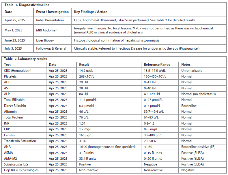
The autoimmune and infectious serology was assessed using indirect immunofluorescence (IIF) for ANA (titer 1:100, homogeneous to fine speckled pattern; laboratory cut-off for positivity: ≥1:80) and enzyme-linked immunosorbent assay (ELISA; Euroimmun AG, Lübeck, Germany) for ASMA and AMA-M2 (cut-offs: >20 R units for positivity). Schistosoma IgG was detected via ELISA (DRG International, Inc., USA; cut-off optical density >1.1), with potential cross-reactivity to other helminths considered but deemed unlikely given the epidemiological context and negative stool studies.
Similarly, for PBC (per AASLD/EASL criteria), AMA-M2 positivity confers high specificity (>95%), but the absence of cholestatic biochemistry (normal ALP, no pruritus or duct lesions) and supportive histology reduces diagnostic probability, suggesting possible false positivity or nonspecific activation rather than overt PBC. Differential considerations included parasitic mimicry versus true overlap syndrome, with biopsy favoring infection.
Given the presence of both infectious and autoimmune markers, ongoing monitoring for potential autoimmune overlap syndromes was considered important in long-term management.
Discussion
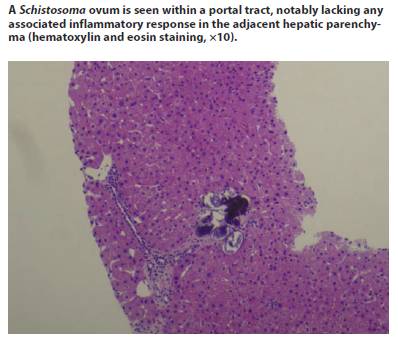
Schistosomiasis is one of the most common human parasitic infections, impacting about 200 million people globally each year [7]. Schistosoma haematobium causes urogenital schistosomiasis, whereas other species lead to gastrointestinal and hepatobiliary schistosomiasis [7]. Schistosomal hepatopathy is usually caused by S. mansoni and, to a lesser degree, by S. japonicum [8]. Humans and other mammals are the main hosts, while freshwater snails act as the intermediate hosts. Cercariae, which are the infective form, are free-swimming larvae that can be found in fresh waters in areas where the disease is common. The cercaria enters the human body by penetrating the skin, travels through the veins, and is transported by the bloodstream to the liver. Adult female and male flatworms develop in the liver, then move to the mesenteric or vesical venous system, where they produce eggs. The eggs are released in the urine or feces, and when they come into contact with fresh water, the life cycle can continue. Eggs are carried by portal blood flow, which helps them get to the hepatic portal microcirculation. The eggs in the liver trigger an initial Th1 immune response, which later develops into a Th2 response. This leads to eosinophilic infiltration, the formation of granulomas, and eventually fibrosis [8, 9]. The final outcome is significant portal fibrosis, which is also referred to as Symmers pipe stem fibrosis. The hepatic lobular architecture is mostly preserved and doesn't have regenerative nodules, which is different from what we see in cirrhosis [6]. However, our patient denied a clear history of freshwater exposure through swimming, fishing, or agricultural work. Additionally, in our patient, histopathology confirmed Schistosomiasis with mild chronic portal inflammation (see Figure), but there was no evidence of granulomas, cirrhosis, or significant portal fibrosis (such as Symmers pipe stem fibrosis), and the hepatic lobular architecture remained preserved.
One notable case described a patient with schistosomiasis who tested positive for ASMA at low titer (1:40) but was initially suspected of autoimmune hepatitis (AIH). Only upon liver biopsy was AIH excluded and schistosomiasis confirmed, with the patient treated effectively with a single dose of praziquantel [9]. In that reported case, both AMA and ANA were negative, in contrast to our patient, who had a broader autoimmune serologic overlap (positive AMA-M2, ASMA, borderline ANA).
Across broader literature, up to 15% of hepatosplenic schistosomiasis cases can exhibit low-titer positivity for autoimmune markers, including ANA, ASMA, or anti-parietal cell antibodies [9]. This mirrors our patient's profile, underscoring that autoimmune seropositivity may reflect nonspecific immune activation secondary to chronic parasitic infection rather than true autoimmune liver disease.
Moreover, whereas some reports highlight portal hypertension or cirrhosis in schistosomiasis [9], our case demonstrated only mild portal inflammation without advanced fibrosis or portal hypertensive sequelae. This marks a milder histopathological course, likely detected early.
Praziquantel treatment, with a single dose of 40 mg/kg, is recommended for patients who have been diagnosed with schistosomiasis. It can be tough to keep track of how well treatment is working. Still, doctor can check the number of eggs being excreted about 4–6 weeks after giving praziquantel, but this only applies to those who were excreting eggs at the start [9]. For our patient, praziquantel, was planned, though details regarding dosage and treatment completion were not available.
Diagnostic pathway and rationale. The diagnosis hinged on integrating epidemiology (previous residence in an endemic area), serology (positive Schistosoma IgG), and crucially, histopathology. The negative stool microscopy, while not uncommon in chronic hepatosplenic disease, underscored the importance of tissue diagnosis. The imaging findings were consistent with chronic liver disease but non-specific. The decision against MRCP was based on the lack of cholestatic clues, making biliary disease a lower probability despite AMA positivity.
Conclusion
In conclusion, this case demonstrates the diagnostic challenge of hepatic schistosomiasis when associated with autoimmune markers of AMA-M2, ASMA and ANA positivity which mimic autoimmune liver disease. Although imaging data were consistent with chronic parenchymal liver disease and elastography consistent with major fibrosis, histology revealed schistosomiasis with only mild chronic portal inflammation and no cirrhosis. Particularly important in reaching the diagnosis was epidemiological history of the patient, including past residence in an endemic area. This case highlights the importance of keeping parasitic infections in the differential diagnosis of chronic liver disease, particularly in people from endemic regions, and the importance of multidisciplinary management including referral to an infectious disease specialist with antiparasitic treatment. Long-term monitoring is still required to evaluate progression of fibrosis and to look for autoimmune overlap.
Conflict of interests. The authors declare that there is not conflict of interests.
Конфликт интересов. Авторы заявляют об отсутствии конфликта интересов.
Financial support. The study was not sponsored (own resources).
Финансирование. Исследование выполнено без спонсорской поддержки (использовались собственные средства).
Authors contributions. Ashraf ALakkad – a major contribution to the development of the concept of the article with writing and editing the case report. Rawia Mubarak Mohamed and Maher Wassim Malaeb – the interpretation of clinical data, critical review of the manuscript. All the authors approved the final version of the article.
Вклад авторов. Ашраф Альаккад – разработка концепции статьи, написание и редактирование описания клинического случая; Равия Мубарак Мохамед и Махер Вассим Малаиб – интерпретация клинических данных, критическая оценка рукописи. Все авторы одобрили окончательную версию статьи для публикации.
Список литературы доступен на сайте журнала https://klin-razbor.ru/
The list of references is available on the journal‘s website https://klin-razbor.ru/
Information about the authors
Информация об авторах
Dr. Rawia Mubarak Mohamed – MD, MBBS, FRCPA Head of Department, Consultant Anatomic Pathology, Burjeel Holdings, UAE. E-mail: rawia.mohamed@burjeelmedicalcity.com;
ORCID: 0009-0000-2320-4063
Равия Мубарак Мохамед – д-р медицины, бакалавр медицины и бакалавр хирургии, член Королевской коллегии патологов Австралии, заведующий кафедрой, консультант по патологической анатомии, Burjeel Holdings, ОАЭ. E-mail: rawia.mohamed@burjeelmedicalcity.com;
ORCID: 0009-0000-2320-4063
Ashraf ALakkad – MD, Internist, Department of Internal Medicine, Chair of Antimicrobial Stewardship Program Madinat Zayed Hospital, UAE. E-mail: ashraf.alaqqad@gmail.com;
ORCID: 0000-0002-4083-2800; Scopus ID: 60052817400; Web Of Science Researcher ID:
AEW-9201-2022
Ашраф Альаккад – д-р медицины, врач-терапевт, кафедра внутренних болезней, руководитель программы управления фармакоэпидемиологическими исследованиями Больницы Мадинат Зайед, ОАЭ. E-mail: ashraf.alaqqad@gmail.com; ORCID: 0000-0002-4083-2800;
Scopus ID: 60052817400; Web Of Science Researcher ID: AEW-9201-2022
Maher Malaeb – MD, Gastroenterologist at Burjeel Royal Hospital Asharej, UAE.
E-mail: maher.malaeb@gmail.com; ORCID: 0000-0002-6415-6595
Махер Малаиб – д-р медицины, гастроэнтеролог, Королевская больница Бурджил, ОАЭ.
E-mail: maher.malaeb@gmail.com; ORCID: 0000-0002-6415-6595
Received: 08.12.2025
Revised: 23.12.2025
Accepted: 29.01.2025
Поступила в редакцию: 08.12.2025
Поступила после рецензирования: 23.12.2025
Принята к публикации: 29.01.2025
Overlapping autoimmune markers in hepatic schistosomiasis: a case report
Rawia Mubarak Mohamed1, Ashraf ALakkad2, Maher Wassim Malaeb1
1 Burjeel Medical City, Abu Dhabi, UAE;
2 Madinat Zayed Hospital, AL Dhafra Region, UAE
ashraf.alaqqad@gmail.com
Abstract
Background. Schistosomiasis is a parasitic infection with hepatobiliary involvement that may mimic autoimmune and chronic liver disease. Its clinical presentation can overlap with conditions such as autoimmune hepatitis and primary biliary cholangitis, particularly in individuals from endemic regions. Diagnosis often requires integration of serology, imaging, and histopathology. We report a case of hepatic schistosomiasis in a middle-aged male presenting with chronic right upper quadrant pain and confounding autoimmune serologies.
Case presentation. A 42-year-old male with no significant past medical or surgical history presented on April 25, 2025, with intermittent nocturnal right upper quadrant (RUQ) abdominal pain and reduced appetite. Initial labs showed preserved liver function (ALT 29 U/L, AST 28 U/L, ALP 84 U/L, total bilirubin 11.4 µmol/L), normal inflammatory markers (CRP 1.7 mg/L), and positive Schistosoma IgG (ELISA assay). Autoimmune workup revealed positive AMA-M2 (33.4 R units) and ASMA (31 R units), with borderline ANA (1:100). Abdominal ultrasound and FibroScan (11.3 kPa) suggested fibrosis; MRI on May 1, 2025, showed irregular liver margins without focal lesions. Stool ova and parasite exam was negative. Liver biopsy on June 23, 2025, confirmed hepatic schistosomiasis with mild chronic portal inflammation. The patient was referred to infectious disease for antiparasitic therapy on July 3, 2025.
Conclusion. This case highlights hepatic schistosomiasis presenting with overlapping autoimmune markers, creating diagnostic complexity. In patients from endemic areas, parasitic infection should remain a differential diagnosis even in the presence of autoimmune antibodies. Long-term follow-up is warranted to monitor for autoimmune overlap and progression of hepatic fibrosis.
Keywords: hepatic schistosomiasis, autoimmune markers, right upper quadrant pain, liver fibrosis, liver biopsy.
For citation: Rawia Mubarak Mohamed, Ashraf ALakkad, Maher Wassim Malaeb. Overlapping autoimmune markers in hepatic schistosomiasis: a case report. Clinical review for general practice. 2025; 7 (6): 103–106. DOI: 10.47407/kr2026.7.7.00893
Introduction
Schistosomiasis, also known as “bilharziasis”, is named after Theodor Bilharzis, who first identified it in 1852 [1]. This tropical disease is caused by trematodes, which are flat-worms from the Schistosoma genus [2]. Schistosomiasis is the second most common human parasitic infection, leading to around 280 thousand deaths each year from complications like renal failure and shock caused by hematemesis [3]. It can lead to a variety of disabilities, including kidney failure, hydronephrosis, hematuria, bladder cancer, and portal hypertension [3]. Schistosomiasis ranks just behind malaria in terms of its impact on public health [4]. Globally, around 200 million people are infected with this parasite, and over 600 million are considered at risk [4]. Right now, there are six known Schistosoma species that can infect humans: S. japonicum, S. haematobium, S. mansoni, S. mekongi, S. intercalatum, and S. malayensis [2]. Infections around the world mainly happen due to the first three species mentioned earlier [2].
Schistosomiasis has various reservoirs besides humans, which complicates efforts to eradicate it in East and Southeast Asia, particularly S. japonicum [5]. Even though schistosomiasis is quite prevalent around the globe, it's pretty uncommon in the United States [6]. However, there are occasional cases, mostly among immigrants from regions where the disease is more widespread [6]. This case presents hepatic schistosomiasis with overlapping autoimmune serologies mimicking autoimmune hepatitis and primary biliary cholangitis.
Case Presentation
A 42-year-old male with no significant past medical or surgical history presented with complaints of intermittent right upper quadrant (RUQ) abdominal pain, which was particularly worse at night. He reported a reduction in appetite but denied nausea, vomiting, or weight loss. His bowel habits were notable for loose stools, without constipation, melena, or hematochezia. There were no urinary complaints, and the patient had never undergone prior gastrointestinal investigations such as esophagogastroduodenoscopy (EGD) or colonoscopy. He denied the use of nonsteroidal anti-inflammatory drugs (NSAIDs). His social history was significant for occasional alcohol consumption in the past, which he stopped one year prior to presentation.
From an epidemiological standpoint, the patient had previously resided in the Philippines before relocating to the UAE, which is relevant given the endemicity of schistosomiasis in certain regions. A clear history of freshwater exposure through swimming, fishing, or agricultural work was not available. He denied any relevant occupational exposures and had no family history of autoimmune liver disease or inflammatory bowel disease. Social and sexual history was noncontributory, as the patient was not sexually active.
A detailed review of systems revealed fatigue but no fever, night sweats, pruritus, jaundice, easy bruising, ascites, or peripheral edema. There was no history of hematemesis, splenomegaly, or variceal bleeding suggestive of portal hypertension. On physical examination, the liver was not palpable, splenomegaly was absent, and no stigmata of chronic liver disease were observed. There was no digital clubbing, and no urinary symptoms were reported despite an enlarged prostate noted on ultrasound.
Diagnostic timeline
The patient's evaluation unfolded as follows (Table 1). Initial presentation occurred on April 25, 2025, with laboratory testing, abdominal ultrasound, and FibroScan. MRI of the liver was performed on May 1, 2025. The patient was initially lost to follow-up but returned for liver biopsy on June 23, 2025. A follow-up visit on July 3, 2025, confirmed clinical stability, with referral to infectious disease for antiparasitic therapy. This sequence underscores the iterative diagnostic process, integrating serology, imaging, and histopathology over approximately 2.5 months.
On initial evaluation dated April 25, 2025, laboratory investigations revealed normal liver function and inflammatory markers, with unremarkable complete blood count and metabolic panel. Detailed laboratory results are summarized in Table 2. Stool ova and parasite examination was performed which was negative for Schistosoma eggs or other parasites; this finding does not exclude chronic infection, as egg excretion may be intermittent or absent in hepatosplenic forms. Specific results included CRP 1.7 mg/L (Ref: 0–5), random glucose 5.5 mmol/L (Ref: 3.89–7.77), albumin 46 g/L (Ref: 39.7–49.4), total protein 76 g/L (Ref: 64–83), ALT 29 U/L (Ref: 0–41), AST 28 U/L (Ref: 0–40), ALP 84 U/L (Ref: 40–129), total bilirubin 11.4 µmol/L (Ref: 0–27), direct bilirubin 6.1 µmol/L, urea 1.98 mmol/L (Ref: 3.2–7.3), creatinine 72 µmol/L (Ref: 62–106), lipase 32 U/L (Ref: 13–60), and INR 1.04. Platelet count was 268×109/L. Thyroid-stimulating hormone and celiac serologies were normal. Stool studies for H. pylori antigen and occult blood were negative, and fecal calprotectin was 9 µg/g (Normal: <50 µg/g).
The autoimmune and infectious serology was assessed using indirect immunofluorescence (IIF) for ANA (titer 1:100, homogeneous to fine speckled pattern; laboratory cut-off for positivity: ≥1:80) and enzyme-linked immunosorbent assay (ELISA; Euroimmun AG, Lübeck, Germany) for ASMA and AMA-M2 (cut-offs: >20 R units for positivity). Schistosoma IgG was detected via ELISA (DRG International, Inc., USA; cut-off optical density >1.1), with potential cross-reactivity to other helminths considered but deemed unlikely given the epidemiological context and negative stool studies.
Similarly, for PBC (per AASLD/EASL criteria), AMA-M2 positivity confers high specificity (>95%), but the absence of cholestatic biochemistry (normal ALP, no pruritus or duct lesions) and supportive histology reduces diagnostic probability, suggesting possible false positivity or nonspecific activation rather than overt PBC. Differential considerations included parasitic mimicry versus true overlap syndrome, with biopsy favoring infection.
Given the presence of both infectious and autoimmune markers, ongoing monitoring for potential autoimmune overlap syndromes was considered important in long-term management.
Discussion
Schistosomiasis is one of the most common human parasitic infections, impacting about 200 million people globally each year [7]. Schistosoma haematobium causes urogenital schistosomiasis, whereas other species lead to gastrointestinal and hepatobiliary schistosomiasis [7]. Schistosomal hepatopathy is usually caused by S. mansoni and, to a lesser degree, by S. japonicum [8]. Humans and other mammals are the main hosts, while freshwater snails act as the intermediate hosts. Cercariae, which are the infective form, are free-swimming larvae that can be found in fresh waters in areas where the disease is common. The cercaria enters the human body by penetrating the skin, travels through the veins, and is transported by the bloodstream to the liver. Adult female and male flatworms develop in the liver, then move to the mesenteric or vesical venous system, where they produce eggs. The eggs are released in the urine or feces, and when they come into contact with fresh water, the life cycle can continue. Eggs are carried by portal blood flow, which helps them get to the hepatic portal microcirculation. The eggs in the liver trigger an initial Th1 immune response, which later develops into a Th2 response. This leads to eosinophilic infiltration, the formation of granulomas, and eventually fibrosis [8, 9]. The final outcome is significant portal fibrosis, which is also referred to as Symmers pipe stem fibrosis. The hepatic lobular architecture is mostly preserved and doesn't have regenerative nodules, which is different from what we see in cirrhosis [6]. However, our patient denied a clear history of freshwater exposure through swimming, fishing, or agricultural work. Additionally, in our patient, histopathology confirmed Schistosomiasis with mild chronic portal inflammation (see Figure), but there was no evidence of granulomas, cirrhosis, or significant portal fibrosis (such as Symmers pipe stem fibrosis), and the hepatic lobular architecture remained preserved.
One notable case described a patient with schistosomiasis who tested positive for ASMA at low titer (1:40) but was initially suspected of autoimmune hepatitis (AIH). Only upon liver biopsy was AIH excluded and schistosomiasis confirmed, with the patient treated effectively with a single dose of praziquantel [9]. In that reported case, both AMA and ANA were negative, in contrast to our patient, who had a broader autoimmune serologic overlap (positive AMA-M2, ASMA, borderline ANA).
Across broader literature, up to 15% of hepatosplenic schistosomiasis cases can exhibit low-titer positivity for autoimmune markers, including ANA, ASMA, or anti-parietal cell antibodies [9]. This mirrors our patient's profile, underscoring that autoimmune seropositivity may reflect nonspecific immune activation secondary to chronic parasitic infection rather than true autoimmune liver disease.
Moreover, whereas some reports highlight portal hypertension or cirrhosis in schistosomiasis [9], our case demonstrated only mild portal inflammation without advanced fibrosis or portal hypertensive sequelae. This marks a milder histopathological course, likely detected early.
Praziquantel treatment, with a single dose of 40 mg/kg, is recommended for patients who have been diagnosed with schistosomiasis. It can be tough to keep track of how well treatment is working. Still, doctor can check the number of eggs being excreted about 4–6 weeks after giving praziquantel, but this only applies to those who were excreting eggs at the start [9]. For our patient, praziquantel, was planned, though details regarding dosage and treatment completion were not available.
Diagnostic pathway and rationale. The diagnosis hinged on integrating epidemiology (previous residence in an endemic area), serology (positive Schistosoma IgG), and crucially, histopathology. The negative stool microscopy, while not uncommon in chronic hepatosplenic disease, underscored the importance of tissue diagnosis. The imaging findings were consistent with chronic liver disease but non-specific. The decision against MRCP was based on the lack of cholestatic clues, making biliary disease a lower probability despite AMA positivity.
Conclusion
In conclusion, this case demonstrates the diagnostic challenge of hepatic schistosomiasis when associated with autoimmune markers of AMA-M2, ASMA and ANA positivity which mimic autoimmune liver disease. Although imaging data were consistent with chronic parenchymal liver disease and elastography consistent with major fibrosis, histology revealed schistosomiasis with only mild chronic portal inflammation and no cirrhosis. Particularly important in reaching the diagnosis was epidemiological history of the patient, including past residence in an endemic area. This case highlights the importance of keeping parasitic infections in the differential diagnosis of chronic liver disease, particularly in people from endemic regions, and the importance of multidisciplinary management including referral to an infectious disease specialist with antiparasitic treatment. Long-term monitoring is still required to evaluate progression of fibrosis and to look for autoimmune overlap.
Conflict of interests. The authors declare that there is not conflict of interests.
Конфликт интересов. Авторы заявляют об отсутствии конфликта интересов.
Financial support. The study was not sponsored (own resources).
Финансирование. Исследование выполнено без спонсорской поддержки (использовались собственные средства).
Authors contributions. Ashraf ALakkad – a major contribution to the development of the concept of the article with writing and editing the case report. Rawia Mubarak Mohamed and Maher Wassim Malaeb – the interpretation of clinical data, critical review of the manuscript. All the authors approved the final version of the article.
Вклад авторов. Ашраф Альаккад – разработка концепции статьи, написание и редактирование описания клинического случая; Равия Мубарак Мохамед и Махер Вассим Малаиб – интерпретация клинических данных, критическая оценка рукописи. Все авторы одобрили окончательную версию статьи для публикации.
Список литературы доступен на сайте журнала https://klin-razbor.ru/
The list of references is available on the journal‘s website https://klin-razbor.ru/
Information about the authors
Информация об авторах
Dr. Rawia Mubarak Mohamed – MD, MBBS, FRCPA Head of Department, Consultant Anatomic Pathology, Burjeel Holdings, UAE. E-mail: rawia.mohamed@burjeelmedicalcity.com;
ORCID: 0009-0000-2320-4063
Равия Мубарак Мохамед – д-р медицины, бакалавр медицины и бакалавр хирургии, член Королевской коллегии патологов Австралии, заведующий кафедрой, консультант по патологической анатомии, Burjeel Holdings, ОАЭ. E-mail: rawia.mohamed@burjeelmedicalcity.com;
ORCID: 0009-0000-2320-4063
Ashraf ALakkad – MD, Internist, Department of Internal Medicine, Chair of Antimicrobial Stewardship Program Madinat Zayed Hospital, UAE. E-mail: ashraf.alaqqad@gmail.com;
ORCID: 0000-0002-4083-2800; Scopus ID: 60052817400; Web Of Science Researcher ID:
AEW-9201-2022
Ашраф Альаккад – д-р медицины, врач-терапевт, кафедра внутренних болезней, руководитель программы управления фармакоэпидемиологическими исследованиями Больницы Мадинат Зайед, ОАЭ. E-mail: ashraf.alaqqad@gmail.com; ORCID: 0000-0002-4083-2800;
Scopus ID: 60052817400; Web Of Science Researcher ID: AEW-9201-2022
Maher Malaeb – MD, Gastroenterologist at Burjeel Royal Hospital Asharej, UAE.
E-mail: maher.malaeb@gmail.com; ORCID: 0000-0002-6415-6595
Махер Малаиб – д-р медицины, гастроэнтеролог, Королевская больница Бурджил, ОАЭ.
E-mail: maher.malaeb@gmail.com; ORCID: 0000-0002-6415-6595
Received: 08.12.2025
Revised: 23.12.2025
Accepted: 29.01.2025
Поступила в редакцию: 08.12.2025
Поступила после рецензирования: 23.12.2025
Принята к публикации: 29.01.2025
Список исп. литературыСкрыть список1. Abdelghani E, Zerpa R, Iliescu G, Escalante CP. Schistosomiasis and liver disease: Learning from the past to understand the present. Clin Case Rep 2020;8(8):1522-6.
2. Rey O, Webster B, Huyse T et al. Population genetics of African Schistosoma species. Infection, Genetics and Evolution 2021;89:104727.
3. Baekby M, Glerup H, Stribolt K, Tarp B. Hepatosplenic schistosomiasis: playing hide-and-seek with an elusive parasite. BMJ case reports 2017;2017.
4. WHO. Current estimated total number of individuals with morbidity and mortality due to Schistosomiasis haematobium and S. mansoni infection in sub-Saharan Africa. 2020.
5. Berger DJ, Léger E, Sankaranarayanan G et al. Genomic evidence of contemporary hybridization between Schistosoma species. PLoS pathogens 2022;18(8):e1010706.
6. Gardner JMF, Mansour NR, Bell AS et al. The discovery of a novel series of compounds with single-dose efficacy against juvenile and adult Schistosoma species. PLOS Neglected Tropical Diseases 2021;15(7):e0009490.
7. Nelwan MLJCTR. Schistosomiasis: life cycle, diagnosis, and control. Current Therapeutic Research 2019;91:5-9.
8. Aula OP, McManus DP, Jones MK, Gordon CA. Schistosomiasis with a focus on Africa. Tropical Medicine and Infectious Disease 2021;6(3):109.
9. Vadukoot Lazar M, Zacharia GS, Shejal AH et al. Hepatic Schistosomiasis: An Overlooked Diagnosis in Non-endemic Regions. Cureus 2025;17(1):e77945.