
Клинический разбор в общей медицине №08 2026
Interleukin-33 as a central immune modulator of glymphatic dysfunction in Alzheimer’s disease: toward a unified immune-fluidic framework
Номера страниц в выпуске:47-51
Abstract
Background. Alzheimer’s disease (AD) remains without effective disease-modifying therapy despite decades of amyloid- and tau-focused interventions. Increasing evidence indicates that diverse pathological processes – including neuroinflammation, astrocytic dysfunction, vascular impairment, and immune dysregulation – converge on a common downstream failure: disruption of glymphatic clearance. However, the immune signals that regulate astrocytic polarity and perivascular fluid transport remain poorly defined.
Aim. To advance a unified immune-fluidic framework positioning interleukin-33 (IL-33) as a central upstream regulator linking neuroimmune signaling to glymphatic dysfunction in AD.
Methods. This narrative review integrates experimental, genetic, biomarker, and translational studies examining IL-33/ST2 signaling, astrocytic aquaporin-4 (AQP4) polarization, microglial reprogramming, and cerebrospinal-interstitial fluid exchange in aging and AD.
Results. IL-33 emerges as a critical regulator of perivascular AQP4 localization rather than total AQP4 expression, thereby governing glymphatic flux and metabolic waste clearance. Experimental IL-33 deficiency leads to astrocytic depolarization, impaired glymphatic drainage, and accelerated amyloid-β and tau accumulation, whereas restoration of IL-33 signaling rescues perivascular architecture and clearance efficiency. IL-33 further orchestrates PU.1-dependent microglial phenotypic shifts, supports mitochondrial and autophagic homeostasis, and operates within a cytokine-competitive signaling network influenced by age, sex, and APOE genotype.
Conclusion. Positioning IL-33 as a master immune-fluidic regulator reframes AD as a disorder of coordinated clearance failure rather than isolated proteinopathy. Therapeutic strategies targeting the IL-33-glymphatic axis may offer disease-modifying potential independent of direct amyloid or tau reduction.
Keywords: interleukin-33, glymphatic system, astrocytic aquaporin-4, neuroimmune regulation, Alzheimer’s disease.
For citation: Abdel-Sater Kh.A. Interleukin-33 as a central immune modulator of glymphatic dysfunction in Alzheimer’s disease: toward a unified immune-fluidic framework. Clinical review for general practice. 2026; 7 (8): 47–51. DOI: 10.47407/kr2026.7.8.00899
Background. Alzheimer’s disease (AD) remains without effective disease-modifying therapy despite decades of amyloid- and tau-focused interventions. Increasing evidence indicates that diverse pathological processes – including neuroinflammation, astrocytic dysfunction, vascular impairment, and immune dysregulation – converge on a common downstream failure: disruption of glymphatic clearance. However, the immune signals that regulate astrocytic polarity and perivascular fluid transport remain poorly defined.
Aim. To advance a unified immune-fluidic framework positioning interleukin-33 (IL-33) as a central upstream regulator linking neuroimmune signaling to glymphatic dysfunction in AD.
Methods. This narrative review integrates experimental, genetic, biomarker, and translational studies examining IL-33/ST2 signaling, astrocytic aquaporin-4 (AQP4) polarization, microglial reprogramming, and cerebrospinal-interstitial fluid exchange in aging and AD.
Results. IL-33 emerges as a critical regulator of perivascular AQP4 localization rather than total AQP4 expression, thereby governing glymphatic flux and metabolic waste clearance. Experimental IL-33 deficiency leads to astrocytic depolarization, impaired glymphatic drainage, and accelerated amyloid-β and tau accumulation, whereas restoration of IL-33 signaling rescues perivascular architecture and clearance efficiency. IL-33 further orchestrates PU.1-dependent microglial phenotypic shifts, supports mitochondrial and autophagic homeostasis, and operates within a cytokine-competitive signaling network influenced by age, sex, and APOE genotype.
Conclusion. Positioning IL-33 as a master immune-fluidic regulator reframes AD as a disorder of coordinated clearance failure rather than isolated proteinopathy. Therapeutic strategies targeting the IL-33-glymphatic axis may offer disease-modifying potential independent of direct amyloid or tau reduction.
Keywords: interleukin-33, glymphatic system, astrocytic aquaporin-4, neuroimmune regulation, Alzheimer’s disease.
For citation: Abdel-Sater Kh.A. Interleukin-33 as a central immune modulator of glymphatic dysfunction in Alzheimer’s disease: toward a unified immune-fluidic framework. Clinical review for general practice. 2026; 7 (8): 47–51. DOI: 10.47407/kr2026.7.8.00899
Интерлейкин-33 как основной иммунный модулятор глимфатической дисфункции при болезни Альцгеймера: к созданию единой иммуножидкостной модели
Х.А. Абдель-СатерУниверситет Мута, Эль-Карак, Иордания
Kabdelsater@mutah.edu.jo
Аннотация
Введение. Для болезни Альцгеймера (БА) по-прежнему не существует эффективной изменяющей течение заболевания терапии, несмотря на десятилетия вмешательств, предполагающих воздействие на амилоид и тау-белок. Все больше данных свидетельствует о том, что разнообразные патологические процессы, в том числе нейровоспаление, дисфункция астроглии, сосудистые нарушения и иммунная дисрегуляция, вместе обусловливают один и тот же сбой: нарушение выведения отходов глимфатической системой. Однако иммунные сигналы, которые регулируют полярность астроцитов и циркуляцию жидкости в периваскулярном пространстве, изучены недостаточно.
Цель. Разработать единую иммуножидкостную модель, в которой интерлейкин-33 (IL-33) рассматривается как основной регулятор, связывающий нейроиммунную сигнализацию с дисфункцией глимфатической системы при БА.
Методы. Настоящий обзор охватывает экспериментальные и генетические исследования, исследования биомаркеров и трансляционные исследования, направленные на изучение сигнализации IL-33/ST2, поляризации астроглиального аквапорина-4 (AQP4), перепрограммирования микроглии и обмена между спинномозговой и интерстициальной жидкостью при старении и БА.
Результаты. IL-33 выступает в качестве важнейшего регулятора периваскулярной локализации AQP4, а не экспрессии AQP4 в целом, обеспечивая регуляцию потока жидкости в глимфатической системе и выведение продуктов метаболизма. Созданный в экспериментальных условиях дефицит IL-33 приводит к деполяризации астроглии, нарушению оттока жидкости в глимфатической системе, а также к усиленному накоплению амилоида-β и тау-белка, в то время как восстановление сигнализации IL-33 обеспечивает восстановление периваскулярной архитектуры и эффективность выведения. IL-33 также регулирует PU.1-зависимые изменения фенотипа микроглии, поддерживает митохондриальный и аутофагальный гомеостаз, функционирует в рамках сети сигналов, конкурирующих с цитокинами, на которую влияют возраст, пол и генотип APOE.
Выводы. Представление об IL-33 как о главном иммуножидкостном регуляторе позволяет пересмотреть взгляды на БА – она скорее является расстройством выведения, чем изолированной протеинопатией. Терапевтические стратегии, предполагающие воздействие на ось IL-33–глимфатическая система, могут изменять течение заболевания независимо от снижения уровня амилоида или тау-белка.
Ключевые слова: интерлейкин-33, глимфатическая система, астроглиальный аквапорин-4, нейроиммунная регуляция, болезнь Альцгеймера.
Для цитирования: Абдель-Сатер Х.А. Интерлейкин-33 как основной иммунный модулятор глимфатической дисфункции при болезни Альцгеймера: к созданию единой иммуножидкостной модели. Клинический разбор в общей медицине. 2026; 7 (8): 47–51. DOI: 10.47407/kr2026.7.8.00899
Introduction
In 2021, an estimated 57 million people worldwide were living with dementia, and over 60% of these cases occurred in low- and middle-income countries. Every year, nearly 10 million new dementia cases are reported globally. Alzheimer’s disease (AD) is the most common form of dementia and may account for 60–70% of all cases [1].
Despite decades of amyloid- and tau-targeted therapies, disease-modifying treatments remain elusive, emphasizing the need to explore upstream, systems-level mechanisms, particularly immune-regulated clearance pathways, that govern protein accumulation and neurodegeneration.
Collectively, these limitations underscore that AD cannot be sufficiently explained by protein-centric models alone and instead demands integrative frameworks that account for dynamic interactions between immune signaling, astrocytic organization, vascular function, and waste clearance.
AD pathogenesis involves interconnected processes: amyloid-β (Aβ) aggregation, tau hyperphosphorylation, microglia-mediated neuroinflammation, blood–brain barrier (BBB) breakdown, vascular dysfunction, oxidative stress, mitochondrial impairment, iron dysregulation, and immune activation [2]. Critically, many of these processes converge on a shared downstream consequence: disruption of glymphatic clearance [3].
The glymphatic system is a perivascular network that circulates cerebrospinal fluid (CSF) and interstitial fluid (ISF) to remove metabolic waste, including Aβ and tau [4]. Aquaporin-4 (AQP4) channels on astrocytic endfeet facilitate convective fluid movement, and their loss or mislocalization reduces waste clearance, accelerating neurodegeneration [5]. Aging, sleep deprivation, vascular stiffening, immune dysregulation, and APOE4 further impair glymphatic activity by reducing arterial pulsation and disrupting AQP4 polarization [4].
Astrocytes, through perivascular endfeet and polarized AQP4, act as both gatekeepers of fluid exchange and immune-responsive cells, positioning them as a critical control point in glymphatic regulation. However, a mechanistic framework linking specific cytokines to AQP4 localization at the gliovascular interface remains lacking [6].
Critically, no mechanistic model has yet defined how immune-derived signals actively instruct astrocytic polarity at the gliovascular interface, despite mounting evidence that astrocytes function as both immune sensors and gatekeepers of glymphatic flux.
Interleukin-33 (IL-33), a member of the IL-1 family, is constitutively expressed in astrocytes and endothelial nuclei and acts as a damage-associated “alarmin”, maintaining immune homeostasis [9]. Human genetic studies identify IL-33 as a susceptibility locus for late-onset AD, with protective polymorphisms such as rs11792633 observed across populations [7].
IL-33 signals through suppression of tumorigenicity 2 (ST2) receptors: membrane-bound ST2L initiates downstream inflammatory signaling via myeloid differentiation primary response 88/ nuclear factor kappa B (MyD88/NF-κB), while soluble ST2 (sST2) acts as a decoy. The ST2L/sST2 balance determines IL-33’s effects [8].
Rather than acting as a downstream consequence of Aβ accumulation, IL-33 functions as a context-dependent coordinator integrating immune tone with astrocytic polarity and perivascular fluid dynamics [9]. This hypothesis yields testable predictions, including that restoration of IL-33 signaling should normalize AQP4 perivascular localization and glymphatic flux even in the absence of direct amyloid reduction, and that disruption of IL-33/ST2 signaling should precede measurable clearance failure.
Consistent with this model, IL-33 deficiency in mice reduces neuron-facing AQP4 (n-AQP4), impairs glymphatic drainage, and accelerates tau accumulation; conversely, recombinant IL-33 restores perivascular AQP4 (p-AQP4) and improves CSF–ISF exchange [10]. This positions IL-33 as a central regulator linking neuroinflammation with glymphatic dysfunction in AD [9].
While prior studies have examined IL-33 biology, glymphatic dysfunction, or astrocytic AQP4 polarity in isolation, no integrative framework has unified these processes into a coherent immune-fluidic axis governing protein clearance in AD. This review addresses that gap by positioning IL-33 as an upstream coordinator linking immune tone to astrocytic architecture and perivascular fluid dynamics.
This review advances a unified immune-fluidic framework for AD, positioning IL-33 as a central regulator that bridges neuroinflammatory signaling with glymphatic clearance dysfunction. We propose that AD-related glymphatic failure stems from a multi-tiered signaling collapse:
1) intracellular competition for shared transducers (e.g., MyD88) blunts IL-33/ST2 efficacy;
2) failed PU.1-mediated microglial state transition impairs waste sensing and phagocytosis;
3) disrupted astrocytic polarity via dystrophin-associated protein complex destabilization compromises convective fluid flow.
Unlike previous compartmentalized approaches, this synthesis integrates IL-33 biology with gliovascular-astrocytic dynamics, offering a novel paradigm in which coordinated intervention at both cytokine-network and fluid-clearance levels is essential for therapeutic success.
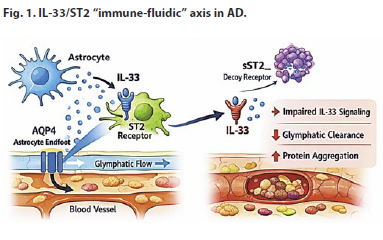
The mechanistic interplay between IL-33 and glymphatic system regulation (Fig. 1)
IL-33 as a central regulator of AQP4 polarization
Experimental studies in Il33⁻/⁻ mice demonstrate a loss of AQP4 from astrocytic endfeet, despite unchanged total AQP4 protein, indicating that IL-33 governs spatial localization rather than expression [10]. IL-33/ST2 signaling stabilizes the dystrophin-associated protein complex and cytoskeletal dynamics, anchoring AQP4 to perivascular membranes [11]. Loss of n-AQP4 impairs neuronal waste clearance, highlighting the primacy of localization over abundance [10].
IL-33 deficiency and glymphatic dysfunction
IL-33 deficiency reduces CSF–ISF exchange, delays solute clearance, and promotes age-dependent tau and Aβ accumulation [12]. Even recombinant IL-33 fails to fully restore neuronal clearance once n-AQP4 polarity is lost, underscoring that perivascular organization is critical for glymphatic efficiency (Fig. 1) [11]. Importantly, post-mortem human studies demonstrating AQP4 depolarization and perivascular astrocytic disruption in AD support the translational relevance of these IL-33-dependent mechanisms observed in animal models.
Astrocyte–Microglia Crosstalk
IL-33 orchestrates PU.1-dependent microglial reprogramming toward a phagocytic, chemotactic phenotype, enhancing debris clearance in parallel with glymphatic drainage [13]. By stabilizing astrocytic cytoskeletons, IL-33 preserves endfoot architecture, maintaining perivascular space patency and facilitating fluid flow [10]. IL-33 signaling efficacy is also modulated by intracellular cytokine competition: pro-inflammatory pathways like NLRP3/IL-1β compete for MyD88, limiting IL-33/ST2 engagement and impairing AQP4 polarization and microglial function [14].
Nevertheless, variability in assay sensitivity, disease stage, and inflammatory context suggests that IL-33 and sST2 should be interpreted as dynamic state markers rather than static diagnostic indicators.
 Integrated neuroprotective effects
IL-33 promotes autophagic flux, mitochondrial health, and reduces oxidative/ER stress and apoptosis [15]. These intracellular effects synergize with glymphatic clearance, creating a positive feedback loop that preserves neuronal integrity and delays AD progression [16].
Conversely, efficient IL-33-dependent glymphatic function prevents accumulation of extracellular neurotoxic species that would otherwise exacerbate neuronal stress. Together, these processes establish a positive feedback loop through which IL-33 preserves neuronal integrity and delays the progression of proteinopathies such as AD [11].
Fig. 1 illustrates a directional, causal model in which IL-33/ST2 signaling stabilizes astrocytic polarity and perivascular AQP4 localization, thereby enabling glymphatic clearance; failure at any node propagates downstream neuroinflammatory amplification and protein accumulation.
IL-33 Dysregulation in AD
Biomarker evidence
AD and mild cognitive impairment (MCI) patients show elevated sST2 and altered IL-33 in CSF/serum, correlating with cognitive decline [17]. Detectable IL-33 is associated with cognitive preservation independent of Aβ or tau, highlighting its neuroprotective potential.
Genetic interactions
Variants in IL-1 receptor-like 1 – IL1RL1 (e.g., rs1921622) modulate sST2 levels and APOE-ε4-associated AD risk, particularly in females, implicating IL-33/ST2 signaling as a modifier of disease susceptibility [18]. APOE4 contributes to microglial dysfunction, cerebrovascular impairment, and oxidative stress, intersecting with IL-33 pathways [19]. These findings reinforce that IL-33/ST2 signaling operates within a genotype- and sex-dependent immunological landscape, with important implications for patient stratification and trial design.
Context-dependent effects
IL-33 exhibits biphasic effects in AD: early-stage IL-33 enhances microglial Aβ clearance and repair, whereas chronic or dysregulated IL-33 signaling may exacerbate neuroinflammation, particularly when sST2 sequesters IL-33 [20].
Therapeutic prospects: targeting IL-33 to restore glymphatic function
Pharmacological strategies
Exogenous recombinant IL-33. Preclinical evidence robustly supports the therapeutic potential of exogenous IL-33. In amyloid precursor protein/presenilin 1 (APP/PS1) transgenic mice, administration of IL-33 Improves microglial phagocytosis, reduces Aβ, restores synaptic function, and promotes anti-inflammatory microglial polarization (Table 1) [21]. These findings collectively demonstrate that IL-33 can enhance glymphatic-associated clearance mechanisms and modulate neuroinflammation, positioning it as a promising disease-modifying candidate. However, therapeutic efficacy is likely contingent on preserved astrocytic architecture; once AQP4 polarity is irreversibly lost, IL-33 supplementation alone may be insufficient, highlighting the importance of early or combination interventions.
ST2 modulation. The IL-33 receptor ST2 mediates both protective and pro-inflammatory signaling in the CNS. Agonists enhance protective IL-33 signaling; inhibitors limit chronic inflammation [18].
Targeting sST2 decoy receptors. Clinical studies show elevated sST2 levels in AD and MCI, where sST2 may sequester IL-33 and attenuate its neuroprotective effects. Targeting sST2 could enhance endogenous IL-33 bioactivity and support glial clearance mechanisms (Table 1) [17].
Gene delivery and engineered cytokines. While not yet applied in AD models, viral or engineered IL-33 constructs with attenuated inflammatory potential are under development in peripheral inflammatory diseases and may be adaptable for CNS-targeted modulation, enabling localized enhancement of astrocyte and microglial functions [22].
Non-pharmacological modulation of IL-33 signaling
Sleep, exercise, and diet. Sleep strongly drives glymphatic clearance, and astrocytic IL-33 expression is integrated with glial responses in homeostasis. Although direct evidence linking lifestyle interventions to IL-33 in humans is emerging, regulatory relationships between IL-33 and astrocyte activity provide a rationale for connecting healthy sleep, physical activity, and metabolic regulation with glymphatic efficiency (see Table 1) [23]. Although causality between lifestyle factors and IL-33 regulation in humans remains to be established, these interventions converge mechanistically on astrocytic health and perivascular dynamics, providing biological plausibility rather than purely associative rationale.
Photobiomodulation & vascular pulsatility. Interventions that enhance cerebrovascular pulsatility and perivascular exchange (e.g., near-infrared photobiomodulation) have been shown to improve glymphatic fluid flow in models of brain injury. Though IL-33 was not directly measured in these studies, its sensitivity to astrocyte and vascular regulation suggests potential synergy with such biophysical approaches (Table 1) [24].
Indirect modulators of the IL-33-glymphatic axis
Microglial phenotype modulators. IL-33 promotes a reparative, phagocytic microglial phenotype, which enhances Aβ and protein clearance. Small molecules or biologics that bias microglia toward this phenotype may potentiate IL-33’s beneficial effects on neural debris removal [21].
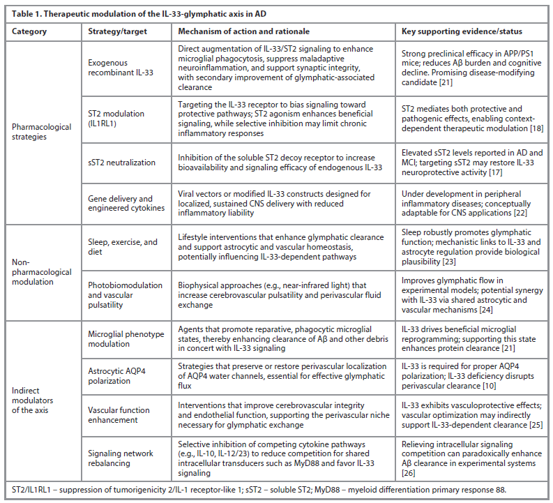
Astrocytic cytoskeleton & AQP4 polarization. IL-33 is required for appropriate AQP4 expression in astrocytes, crucial for glymphatic flux. IL-33 deficiency reduces astrocytic AQP4, impairing perivascular water transport and clearance of pathogenic proteins [10].
Vascular function enhancers. IL-33 has vasculoprotective roles and may contribute to endothelial repair pathways that support glymphatic exchange. Although specific studies in AD are limited, enhancing cerebrovascular health remains a viable route to support IL-33-dependent clearance mechanisms (Table 1) [25].
Signaling network rebalancing. Experimental evidence indicates that inhibiting certain cytokines – such as IL-10 or IL-12/IL-23 – can paradoxically enhance Aβ clearance by relieving competition for shared intracellular transducers (e.g., MyD88) and thereby 'unblocking' beneficial IL-33 signaling. This suggests the IL-33/ST2/AQP4 axis operates within a push-pull immunoregulatory network [26].
Despite promising mechanistic rationale, several barriers persist:
• Safety: IL-33’s dual anti- and pro-inflammatory roles require careful dosing and timing optimization to avoid exacerbating neuroinflammation [20].
• Aging effects: age-related changes in astrocytes and IL-33 responsiveness may limit efficacy in late-stage AD [27].
• Blood–brain barrier delivery: efficient and targeted delivery of IL-33–based therapies into specific brain regions remains a key obstacle [28].
• Biomarker integration: joint profiling of CSF IL-33/ sST2 with advanced glymphatic imaging could serve as biomarkers of engagement, but standardized protocols are lacking [29].
Patient stratification – including sex, APOE genotype, and disease stage – will be essential for IL-33-targeted clinical trials, given known variability in immune responsiveness, astrocytic polarity, and glymphatic function across these dimensions [30].
Critical evaluation and limitations
The IL-33/ST2 signaling axis is increasingly associated with AD, with clinical studies reporting reduced IL-33 and elevated sST2 in serum and CSF of individuals with MCI and AD compared with healthy controls, consistent with dysregulated IL-33/ST2 signaling in dementia [31]. Meta analytic evidence also indicates significant relationships between IL-33/ST2 levels and AD susceptibility, though findings vary across cohorts and methodologies [18].
Genetic polymorphisms in IL-33/ST2 genes further suggest modulation of disease risk and potential interaction with APOE4 status, but replication in larger and more diverse populations is necessary to confirm these associations [32].
Mechanistic insights into IL-33’s role in AD derive largely from preclinical animal and cellular models, which do not fully capture human neuroinflammation, cerebrovascular aging, glymphatic dynamics, and the heterogeneity of late-onset AD. Rodent models recapitulate amyloid pathology but lack the full complexity of human chronic inflammation and multi factorial risk factors. Evidence linking glymphatic dysfunction to AD in humans remains limited, with clinical imaging and longitudinal assessments still emerging and no standardized in vivo measures widely adopted [33]. Integration of IL-33/sST2 profiling with emerging glymphatic imaging modalities may enable functional stratification of clearance capacity, but such multimodal approaches require harmonized protocols and longitudinal validation.
Human studies are further constrained by cross-sectional designs, small sample sizes, and variability in IL-33 and sST2 assay methods, which often lack the sensitivity to distinguish active versus inactive cytokine forms, complicating interpretation of results [34]. Longitudinal investigation of IL-33/ ST2 dynamics in relation to cognitive decline, glymphatic function, sleep-dependent CSF clearance, astrocyte heterogeneity, and regional brain vulnerability remains sparse [35].
Finally, IL-33 exhibits context dependent effects in AD; it can support neuroprotective processes under some conditions, but its signaling may be attenuated or counterproductive when sST2 levels are high or in the setting of chronic inflammation. This complexity emphasizes the need for precision based, stage specific approaches, including stratification by disease stage, APOE genotype, sex, and immune profile, to optimize biomarker development and design targeted therapeutic trials [36].
Conclusion and future perspectives
IL-33 serves as a central coordinator of glymphatic and neuroimmune homeostasis in AD, regulating AQP4 polarization, supporting astrocytic integrity, and orchestrating microglial clearance. Therapeutic strategies enhancing IL-33 signaling – pharmacological, gene-based, or lifestyle-mediated – may slow neurodegeneration independent of direct Aβ or tau reduction. Critical questions remain regarding IL-33 regulation of AQP4 polarity, interactions with sleep, vascular dynamics, APOE genotype, and safe CNS delivery. Positioning IL-33 as an immune-fluidic regulator reframes AD as a disorder of coordinated clearance failure, suggesting that successful disease modification will require synchronizing immunomodulation with restoration of astrocytic and vascular architecture rather than targeting protein aggregates alone.
Conflict of interests. The author declares that there is not conflict of interests.
Конфликт интересов. Автор заявляет об отсутствии конфликта интересов.
Funding. This research received no external funding.
Финансирование. Исследование проведено без финансовой поддержки.
Список литературы доступен на сайте журнала https://klin-razbor.ru/
The list of references is available on the journal‘s website https://klin-razbor.ru/
Information about the author
Информация об авторе
Khaled A. Abdel-Sater – MD, Faculty of Dentistry, Mutah University.
E-mail: Kabdelsater@mutah.edu.jo; ORCID: 0000-0001-9357-4983
Халед А. Абдель-Cатер – доктор медицины, стоматологический факультет, Университет Мута. E-mail: Kabdelsater@mutah.edu.jo; ORCID: 0000-0001-9357-4983
Received: 02.03.2026
Revised: 05.03.2026
Accepted: 12.03.2026
Поступила в редакцию: 02.03.2026
Поступила после рецензирования: 05.03.2026
Принята к публикации: 12.03.2026
Список исп. литературыСкрыть список1. World Health Organization. Dementia: Key Facts. 31 March 2025. https://www.who.int/news-room/fact-sheets/detail/dementia
2. Zhang J, Kong G, Yang J et al. Pathological mechanisms and treatment progression of Alzheimer's disease. Eur J Med Res 2025;30(1):625. DOI: 10.1186/s40001-025-02886-9
3. Chen Y, He Y, Han J et al. Blood-brain barrier dysfunction and Alzheimer's disease: associations, pathogenic mechanisms, and therapeutic potential. Front Aging Neurosci 2023;(15):1258640. DOI: 10.3389/fnagi.2023.1258640
4. Silva I, Silva J, Ferreira R, Trigo D. Glymphatic system, AQP4, and their implications in Alzheimer's disease. Neurol Res Pract 2021;3(1):5. DOI: 10.1186/s42466-021-00102-7
5. Mohaupt P, Vialaret J, Hirtz C, Lehmann S. Readthrough isoform of aquaporin-4 (AQP4) as a therapeutic target for Alzheimer's disease and other proteinopathies. Alzheimers Res Ther 2023;15(1):170. DOI: 10.1186/s13195-023-01318-2
6. Salman MM, Kitchen P, Halsey A et al. Emerging roles for dynamic aquaporin-4 subcellular relocalization in CNS water homeostasis. Brain 2022;145(1):64-75. DOI: 10.1093/brain/awab311
7. Sheng F, Li M, Yu JM et al. IL-33/ST2 axis in diverse diseases: regulatory mechanisms and therapeutic potential. Front Immunol 2025;(16):1533335. DOI: 10.3389/fimmu.2025.1533335
8. Hawerkamp HC, Yeow A, Byrne CM et al. The interleukin-33 receptor (ST2) is a novel therapeutic target to attenuate the progression of hemophilic arthropathy. Blood 2025;146(16):1979-90. DOI: 10.1182/blood.2024027578
9. Cayrol C, Girard JP. Interleukin-33 (IL-33): A critical review of its biology and the mechanisms involved in its release as a potent extracellular cytokine. Cytokine 2022;(156):155891. DOI: 10.1016/j.cyto.2022.155891
10. Wu J, Carlock C, Shim J et al. Requirement of brain interleukin33 for aquaporin4 expression in astrocytes and glymphatic drainage of abnormal tau. Mol Psychiatry 2021;26(10):5912-24. DOI: 10.1038/s41380-020-00992-0
11. Liu Y, Hu PP, Zhai S et al. Aquaporin 4 deficiency eliminates the beneficial effects of voluntary exercise in a mouse model of Alzheimer's disease. Neural Regen Res 2022;17(9):2079-88. DOI: 10.4103/1673-5374.335169
12. Gomolka RS, Hablitz LM, Mestre H et al. Loss of aquaporin-4 results in glymphatic system dysfunction via brain-wide interstitial fluid stagnation. Elife 2023;(12):e82232.
13. Vainchtein ID, Chin G, Cho FS et al. Astrocyte-derived interleukin-33 promotes microglial synapse engulfment and neural circuit development. Science 2018;359(6381):1269-73. DOI: 10.1126/science.aal3589
14. Zharichenko N, Njoku DB. The Role of Pro-Inflammatory and Regulatory Signaling by IL-33 in the Brain and Liver: A Focused Systematic Review of Mouse and Human Data and Risk of Bias Assessment of the Literature. Int J Mol Sci 2020;21(11):3933. DOI: 10.3390/ijms21113933
15. Xie D, Liu H, Xu F et al. IL33 (Interleukin 33)/ST2 (Interleukin 1 Receptor-Like 1) Axis Drives Protective Microglial Responses and Promotes White Matter Integrity After Stroke. Stroke 2021;52(6):2150-61. DOI: 10.1161/STROKEAHA.120.032444
16. Xu R, He Q, Wang Y et al. Therapeutic Potential of Remote Ischemic Conditioning in Vascular Cognitive Impairment. Front Cell Neurosci 2021;(15):706759. DOI: 10.3389/fncel.2021.706759
17. Saresella M, Marventano I, Piancone F et al. IL-33 and its decoy sST2 in patients with Alzheimer's disease and mild cognitive impairment. J Neuroinflammation 2020;17(1):174. DOI: 10.1186/s12974-020-01806-4
18. Jiang Y, Zhou X, Wong HY et al. An IL1RL1 genetic variant lowers soluble ST2 levels and the risk effects of APOE-ε4 in female patients with Alzheimer's disease. Nat Aging 2022;2(7):616-34. DOI: 10.1038/s43587-022-00241-9
19. Pires M, Rego AC. Apoe4 and Alzheimer's Disease Pathogenesis-Mitochondrial Deregulation and Targeted Therapeutic Strategies. Int J Mol Sci 2023;24(1):778. DOI: 10.3390/ijms24010778
20. Rao X, Hua F, Zhang L et al. Dual roles of interleukin-33 in cognitive function by regulating central nervous system inflammation. J Transl Med 2022;20(1):369. DOI: 10.1186/s12967-022-03570-w
21. Fu AK, Hung KW, Yuen MY et al. IL-33 ameliorates Alzheimer's disease-like pathology and cognitive decline. Proc Natl Acad Sci U S A 2016;113(19):E2705-E2713. DOI: 10.1073/pnas.1604032113
22. Zhang H, Chen H, Hu X et al. Inflammation-modulating polymeric nanoparticles: design strategies, mechanisms, and therapeutic applications. EBioMedicine 2025;(118):105837. DOI: 10.1016/j.ebiom.2025.105837
23. Chong PLH, Garic D, Shen MD et al. Sleep, cerebrospinal fluid, and the glymphatic system: A systematic review. Sleep Med Rev 2022;(61):101572. DOI: 10.1016/j.smrv.2021.101572
24. Lopes DM, Wells JA, Ma D et al. Glymphatic inhibition exacerbates tau propagation in an Alzheimer's disease model. Alzheimers Res Ther 2024;16(1):71. DOI: 10.1186/s13195-024-01439-2
25. Liu M, Huang J, Liu T et al. Exogenous interleukin 33 enhances the brain's lymphatic drainage and toxic protein clearance in acute traumatic brain injury mice. Acta Neuropathol Commun 2023;11(1):61. DOI: 10.1186/s40478-023-01555-4
26. Krajewski D, Ranjitkar S, Jordan N et al. IL-33 signaling is dispensable for the IL-10-induced enhancement of mast cell responses during food allergy. Front Immunol 2025;(16):1526498. DOI: 10.3389/fimmu.2025.1526498
27. Lou Y. Role of Interleukin33 in Rejuvenation of Aged Neurons and Age-Related Dementias. Neurosci Insights 2021;(16):26331055211030251. DOI: 10.1177/26331055211030251
28. Wu D, Chen Q, Chen X et al. The blood-brain barrier: structure, regulation, and drug delivery. Signal Transduct Target Ther 2023;8(1):217. DOI: 10.1038/s41392-023-01481-w
29. Firouzabadi N, Dashti M, Dehshahri A, Bahramali E. Biomarkers of IL-33 and sST2 and Lack of Association with Carvedilol Therapy in Heart Failure. Clin Pharmacol 2020;(12):53-8. DOI: 10.2147/CPAA.S256290
30. Murdaca G, Paladin F, Tonacci A et al. Involvement of IL-33 in the Pathogenesis and Prognosis of Major Respiratory Viral Infections: Future Perspectives for Personalized Therapy. Biomedicines 2022;10(3):715. DOI: 10.3390/biomedicines10030715
31. Jiang T, Zheng T, Li W et al. IL-33/ST2 signaling pathway and Alzheimer's disease: A systematic review and meta-analysis. Clin Neurol Neurosurg 2023;(230):107773. DOI: 10.1016/j.clineuro.2023.107773
32. Pei M, Liu X, Yang P et al. Genetic Association of Interleukin 33/ST2 Polymorphisms With Behcet's Uveitis. Front Immunol 2021;(12):589639. DOI: 10.3389/fimmu.2021.589639
33. Keil SA, Jansson D, Braun M, Iliff JJ. Glymphatic dysfunction in Alzheimer's disease: A critical appraisal. Science 2025;389(6756):eadv8269. DOI: 10.1126/science.adv8269
34. Liang CS, Su KP, Tsai CL et al. The role of interleukin-33 in patients with mild cognitive impairment and Alzheimer's disease. Alzheimers Res Ther 2020;12(1):86. DOI: 10.1186/s13195-020-00652-z
35. Sharkey RJ, Cortese F, Goodyear BG et al. Longitudinal analysis of glymphatic function in amyotrophic lateral sclerosis and primary lateral sclerosis. Brain 2024;147(12):4026-32. DOI: 10.1093/brain/awae288
36. Chen T, Chen J, Guo M et al. IL-33 exerts neuroprotective effects through activation of ST2/AKT signaling axis in microglia after subarachnoid hemorrhage in rats. Neuropharmacology 2025;(269):110336. DOI: 10.1016/j.neuropharm.2025.110336