
Клинический разбор в общей медицине №08 2026
Hyperactive delirium following left basal ganglia intracerebral hemorrhage: a neuropsychiatric case report
Номера страниц в выпуске:67-69
Abstract
Spontaneous intracerebral hemorrhage is frequently complicated by neuropsychiatric disturbances, including delirium, which remains underrecognized despite its association with poor clinical outcomes. We report a case of a 44-year-old man with untreated hypertension who presented with acute right-sided weakness, dysarthria, and fluctuating consciousness. Neuroimaging revealed a left basal ganglia intracerebral hemorrhage measuring 8.77 cc, accompanied by perifocal edema and mild midline shift. During hospitalization, the patient developed hyperactive delirium characterized by agitation, irritability, disorientation, and a sundowning pattern. Mental status examination demonstrated impaired attention, fluctuating awareness, and affective dysregulation, with no prior psychiatric history. The diagnosis of delirium due to another medical condition (ICD-10: F05) was established based on clinical features and the Confusion Assessment Method for the ICU. Management included low-dose haloperidol and non-pharmacological interventions such as environmental reorientation and sleep–wake cycle regulation. This case highlights the neuropsychiatric impact of subcortical injury involving the basal ganglia and underscores the importance of early recognition and multidisciplinary management, including consultation liaison psychiatry, to optimize patient safety and functional outcomes.
Keywords: intracerebral hemorrhage, basal ganglia, delirium, neuropsychiatry, consultation liaison psychiatry.
For citation: Rini I.W.A., Maramis M.M., Haryono Y., Yulianti E. Hyperactive delirium following left basal ganglia intracerebral hemorrhage: a neuropsychiatric case report. Clinical review for general practice. 2026; 7 (8): 67–69. DOI: 10.47407/kr2026.7.8.00904
Spontaneous intracerebral hemorrhage is frequently complicated by neuropsychiatric disturbances, including delirium, which remains underrecognized despite its association with poor clinical outcomes. We report a case of a 44-year-old man with untreated hypertension who presented with acute right-sided weakness, dysarthria, and fluctuating consciousness. Neuroimaging revealed a left basal ganglia intracerebral hemorrhage measuring 8.77 cc, accompanied by perifocal edema and mild midline shift. During hospitalization, the patient developed hyperactive delirium characterized by agitation, irritability, disorientation, and a sundowning pattern. Mental status examination demonstrated impaired attention, fluctuating awareness, and affective dysregulation, with no prior psychiatric history. The diagnosis of delirium due to another medical condition (ICD-10: F05) was established based on clinical features and the Confusion Assessment Method for the ICU. Management included low-dose haloperidol and non-pharmacological interventions such as environmental reorientation and sleep–wake cycle regulation. This case highlights the neuropsychiatric impact of subcortical injury involving the basal ganglia and underscores the importance of early recognition and multidisciplinary management, including consultation liaison psychiatry, to optimize patient safety and functional outcomes.
Keywords: intracerebral hemorrhage, basal ganglia, delirium, neuropsychiatry, consultation liaison psychiatry.
For citation: Rini I.W.A., Maramis M.M., Haryono Y., Yulianti E. Hyperactive delirium following left basal ganglia intracerebral hemorrhage: a neuropsychiatric case report. Clinical review for general practice. 2026; 7 (8): 67–69. DOI: 10.47407/kr2026.7.8.00904
Гиперактивный делирий после внутримозгового кровоизлияния в базальные ядра слева: случай нейропсихиатрического расстройства
И.В.А. Рини, М.М. Марамис, Ю. Харионо, Э. Юлианти1 Университет Айрлангга, Сурабая, Индонезия;
2 Университетская больница общего профиля им. доктора Соетомо, Сурабая, Индонезия
margarit@fk.unair.ac.id
Аннотация
Осложнениями спонтанного внутримозгового кровоизлияния часто бывают нейропсихиатрические расстройства, в том числе делирий, который остается недостаточно изученным, несмотря на его связь с неблагоприятными клиническими исходами. Представлено описание случая 44-летнего мужчины с нелеченой артериальной гипертензией, обратившегося с жалобами на внезапно возникшую слабость в правой половине тела, дизартрию и нарушение сознания. Нейровизуализационное исследование выявило внутримозговое кровоизлияние объемом 8,77 см3 в базальные ядра слева, сопровождающееся перифокальным отеком и небольшим смещением срединных структур. Во время пребывания в стационаре у пациента развился гиперактивный делирий, характеризовавшийся наличием возбуждения, раздражительности, дезориентации и синдрома заката. Оценка психического состояния выявила нарушение внимания, нарушение сознания и эмоциональную дисрегуляцию при отсутствии психических расстройств в анамнезе. Диагноз «делирий, не вызванный алкоголем или другими психоактивными веществами» (F05 по Международной классификации болезней 10-го пересмотра) поставлен на основании клинических проявлений и результатов применения метода оценки спутанности сознания для отделения реанимации и интенсивной терапии (CAM-ICU). Лечение предполагало использование низких доз галоперидола и немедикаментозные вмешательства, такие как восстановление ориентации в окружающем мире и регуляция цикла сна и бодрствования. Представленный случай акцентирует внимание на нейропсихиатрических последствиях повреждения подкорковых структур, затрагивающего базальные ядра, и подчеркивает важность раннего выявления и мультидисциплинарного подхода к лечению, в том числе предполагающего консультацию психиатра, для повышения безопасности пациентов и улучшения функциональных исходов.
Ключевые слова: внутримозговое кровоизлияние, базальные ядра, делирий, нейропсихиатрия, консультативная психиатрия.
Для цитирования: Рини И.В.А., Марамис М.М., Харионо Ю., Юлианти Э. Гиперактивный делирий после внутримозгового кровоизлияния в базальные ядра слева: случай нейропсихиатрического расстройства. Клинический разбор в общей медицине. 2026; 7 (8): 67–69. DOI: 10.47407/kr2026.7.8.00904
Introduction
Spontaneous intracerebral hemorrhage (ICH) represents a critical intersection between neurology and psychiatry, in which structural brain injury frequently precipitates neuropsychiatric syndromes. Although acute management primarily focuses on hemodynamic stabilization and neurosurgical evaluation, delirium remains a common but frequently underrecognized complication. Delirium in ICH is associated with prolonged hospitalization, functional dependency, and increased mortality [1, 2].
Delirium is defined as an acute and fluctuating disturbance of attention, awareness, and cognition resulting from multifactorial cerebral dysfunction. In neurological settings, delirium is often misattributed to nonspecific agitation, particularly when focal neurological deficits interfere with cognitive assessment. This diagnostic challenge is especially relevant in basal ganglia hemorrhage, where disruption of cortico-striato-thalamo-cortical circuits affects not only motor function but also executive and affective regulation [3].
Consultation liaison psychiatry (CLP) offers a structured approach for identifying and managing neuropsychiatric complications in medically ill patients. Early psychiatric involvement supports timely diagnosis, symptom control, and interdisciplinary coordination, which are essential in acute stroke care.
We report a case of hyperactive delirium in a middle-aged male with unilateral left basal ganglia ICH, highlighting the neuropsychiatric consequences of subcortical injury and the clinical value of early CLP intervention.
Case presentation
Mr. H, a 44-year-old male with a history of untreated hypertension, presented with sudden onset of right-sided numbness and weakness shortly after leaving the bathroom. Symptoms rapidly progressed to dysarthria and fluctuating consciousness. He was initially treated at a local hospital before referral to a tertiary care center.
During hospitalization, the patient developed prominent behavioral disturbances, including agitation, irritability, and psychomotor restlessness, with marked evening exacerbation consistent with a sundowning pattern. He repeatedly attempted to remove medical devices, necessitating close supervision and brief use of physical restraints for safety.
The patient remained disoriented to place, intermittently believing he was at home, and demonstrated perseverative thoughts related to returning to his work crafting traditional musical instruments. On admission, he was in hypertensive crisis. Neurological examination revealed right-sided hemiparesis and cranial nerve deficits consistent with an upper motor neuron lesion.
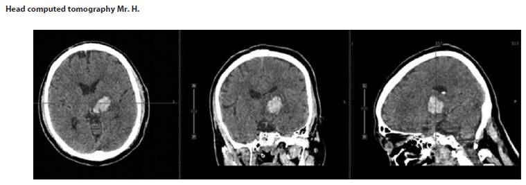
Non-contrast head computed tomography demonstrated a left basal ganglia ICH with an estimated volume of 8.77 cc, associated with perifocal edema, mild compression of the left lateral ventricle, and a 2.1 mm midline shift. Laboratory findings showed leukocytosis, elevated fibrinogen and D-dimer levels, hyperuricemia, and hypertriglyceridemia, without evidence of systemic infection (see Figure).
Mental status examination revealed fluctuating consciousness, impaired attention, disorientation, dysphoric mood, and intermittent inappropriate smiling suggestive of affective dysregulation. No prior psychiatric history was reported. Based on clinical features and CAM-ICU findings, the patient was diagnosed with delirium due to another medical condition (acute ICH; ICD-10: F05). Premorbid collateral history suggested rigid and meticulous personality traits, which were considered descriptive rather than diagnostic.
Clinical assessment. Serial neurological and psychiatric assessments were conducted during hospitalization. Standardized instruments included the Confusion Assessment Method for the ICU (CAM-ICU), Richmond Agitation Sedation Scale (RASS), National Institutes of Health Stroke Scale (NIHSS), Intracerebral Hemorrhage (ICH) Score, secondary ICH (sICH) Score, and the Modified Rankin Scale (mRS).
Management. Behavioral disturbances were managed with low-dose haloperidol (0.5 mg orally twice daily), chosen for its efficacy in controlling hyperactive delirium while allowing ongoing neurological monitoring. Non-pharmacological interventions included environmental reorientation, sleep-wake cycle regulation, and family involvement.
Discussion
The basal ganglia play a central role in integrating motor, cognitive, and affective processes through extensive cortico-subcortical networks. In this case, unilateral basal ganglia hemorrhage resulted in prominent delirium despite relative cortical preservation. This observation aligns with contemporary models conceptualizing delirium as a disorder of large-scale brain network connectivity, particularly involving subcortical hubs regulating arousal and attention [4].
From a neuroanatomical perspective, the basal ganglia form the core of cortico-striato-thalamo-cortical circuits supporting executive control and motivational regulation. Disruption of these pathways may impair attentional integration and emotional modulation, precipitating delirium even in the absence of widespread cortical injury [3, 5]. Functional neuroimaging studies further support this mechanism, demonstrating reduced subcortical connectivity and impaired interaction between executive networks and the default mode network in delirium [4].
Delirium is a frequent but underrecognized complication of ICH. А. Naidech et al. reported delirium symptoms in approximately 27% of patients with ICH, with significant associations with longer hospital stays, poorer functional outcomes, and reduced quality of life [6]. More recent cohort data indicate that delirium may occur in nearly 60% of ICH patients, and that delirium resolution prior to discharge is associated with improved functional outcomes [7]. These findings underscore the diagnostic challenges of delirium in the presence of focal neurological deficits.
Laterality and lesion characteristics have important prognostic implications. Bilateral basal ganglia hemorrhage is associated with severe impairment of consciousness and poor outcomes, whereas unilateral lesions generally show a more favorable trajectory [8]. Nonetheless, lesion location and hematoma volume remain key determinants of outcome [9].
Perihematomal edema has emerged as an additional prognostic factor. Increased perihematomal edema volume and rapid expansion are associated with worse functional outcomes, particularly in basal ganglia hemorrhage, even when hematoma size is relatively small [10].
Early CLP involvement in this case aligns with current recommendations emphasizing multidomain delirium management, including active detection, environmental optimization, and interdisciplinary coordination. Routine delirium screening using tools such as the CAM-ICU facilitates early identification and may improve clinical outcomes in ICH patients [4, 6].
Conclusion
This case illustrates the neuropsychiatric consequences of unilateral basal ICH and highlights delirium as a critical marker of acute brain dysfunction. Early recognition and management through CLP may improve patient safety, support neurological assessment, and facilitate functional recovery. These findings should be interpreted within the limitations of a single-case report.
Conflict of interests. The authors declare that there is not conflict of interests.
Конфликт интересов. Авторы заявляют об отсутствии конфликта интересов.
Ethics approval and patient consent. Ethical approval was obtained from the appropriate institutional review board. Written informed consent was obtained from the patient for publication of this case report and accompanying clinical information.
Соблюдение этических стандартов и согласие пациента. Получено одобрение этического комитета соответствующего учреждения. От пациента получено письменное добровольное информированное согласие на публикацию данного клинического случая и соответствующих клинических данных.
Список литературы доступен на сайте журнала https://klin-razbor.ru/
The list of references is available on the journal‘s website https://klin-razbor.ru/
Information about the authors
Информация об авторах
Ika Wahyu Adita Rini – Psychiatric Resident, Department of Psychiatry, Faculty of Medicine, Universitas Airlangga, Dr. Soetomo General Academic Hospital
Рини Ика Вахью Адита – ординатор психиатрического отд-ния, каф. психиатрии, медицинский фак-т, Университет Айрлангга, Университетская больница общего профиля им. доктора Соетомо
Margarita Maria Maramis – Prof. Dr. dr. Sp.KJ (K), Department of Psychiatry, Faculty of Medicine, Universitas Airlangga, Dr. Soetomo General Academic Hospital. E-mail: margarit@fk.unair.ac.id
Марамис Маргарита Мария – проф., доктор медицины, врач-психиатр (консультирующий), каф. психиатрии, медицинский фак-т, Университет Айрлангга, Университетская больница общего профиля им. доктора Соетомо. E-mail: margarit@fk.unair.ac.id
Yudha Haryono – Dr. dr. Sp.N, Department of Neurology, Faculty of Medicine, Universitas Airlangga
Юдха Харионо – доктор медицины, врач-невролог, каф. неврологии, медицинский фак-т, Университет Айрлангга, Университетская больница общего профиля им. доктора Соетомо
Erikavitri Yulianti – dr. Sp.KJ (K), Department of Psychiatry, Faculty of Medicine, Universitas Airlangga, Dr. Soetomo General Academic Hospital
Эрикавитри Юлианти – врач-психиатр (консультирующий), каф. психиатрии, медицинский фак-т, Университет Айрлангга, Университетская больница общего профиля им. доктора Соетомо
Received: 30.03.2026
Revised: 10.04.2026
Accepted: 16.04.2026
Поступила в редакцию: 30.03.2026
Поступила после рецензирования: 10.04.2026
Принята к публикации: 16.04.2026
Список исп. литературыСкрыть список1. Reznik ME, Drake J, Margolis SA et al. Delirium in neurocritical care. Crit Care Med 2019;47(10):1360-8.
2. Marrama F, Kyheng M, Pasi M et al. Neuropsychiatric complications of intracerebral hemorrhage. Int J Stroke 2022;17(2):158-70.
3. Murphy S, Werring D. Stroke: causes and clinical features. Medicine (UK) 2020;48(10):620-6.
4. Wilson JE, Mart MF, Cunningham C et al. Delirium. Nat Rev Dis Primers 2020;(6):90.
5. Wichmann T, DeLong MR. Functional and pathophysiological models of the basal ganglia. Curr Opin Neurobiol 1996;6(6):751-8.
6. Naidech AM, Beaumont JL, Rosenberg NF et al. Intracerebral hemorrhage and delirium symptoms: length of stay, function, and quality of life. Am J Respir Crit Care Med 2013;188(11):1331-7.
7. Reznik ME, Margolis SA, Mahta A et al. Impact of delirium on outcomes after intracerebral hemorrhage. Stroke 2022;53(2):505-13.
8. Watanabe G, Conching AK, Ogasawara CT et al. Bilateral basal ganglia hemorrhage: a systematic review. Neurosurg Rev 2023;(46):170.
9. Park J, Jang H. Association between hemorrhage location and prognosis in spontaneous intracerebral hemorrhage of the basal ganglia and thalamus. Medicine (Baltimore) 2022;101(39):e32000.
10. Murthy SB, Moradiya Y, Dawson J et al. Perihematomal edema and functional outcomes in intracerebral hemorrhage. Stroke 2016;47(Suppl.1):WMP94.
29 сентября 2026
Количество просмотров: 100